

Growth morphology on PDA of Phytophthora lateralis
Photo Gallery
|
P. lateralis colony morphology on PDA 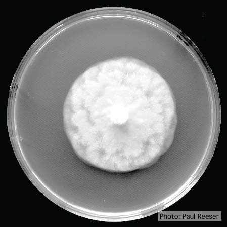 |
P. pluvialis on Pinus radiata in New Zealand  Pinus radiata needles showing colour changes following infection with red needle cast disease. The tissues around the initial infection at the base or along the needle senesce, and change yellow and then brown as indicated by the arrows before the needles cast. |
Growth of P. arenaria on MEA 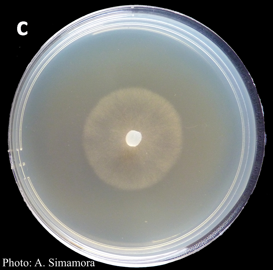 Colony morphology of Phytophthora arenaria after 7 days at 20°C on malt extract agar |
|
P. austrocedrae hyphal swellings in liquid media drawing  Morphology of hyphae of Phytophthora austrocedrae, from Greslebin et al. 2007 |
P. cinnamomi on Jarrah 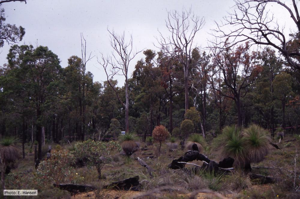 Dieback in Jarrah, Western Australia |
P. nicotianae colony morphology on PDA 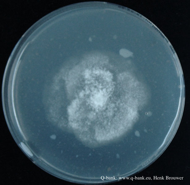 Phytophthora nicotianae CBS 321.49 PDA after 7 days at 24 degrees. Photo from Q-bank: www.q-bank.eu, Henk Brouwer (CBS-KNAW, Utrecht, The Netherlands) |
|
P. megasperma colony morphology on V8  Colony morphology on V8 at 7 days |
P. agathidicida oospores  Oospores of P. agathidicida in the roots of kauri seedlings inoculated with P. agathidicida. The root has been cleared with potassium hydroxide and bleached with peroxide, before being stained with Trypan Blue |
P. tentaculata chlamydospore  P. tentaculata chlamydospore with short hyphal projection |
|
P. nicotianae chlamydospore 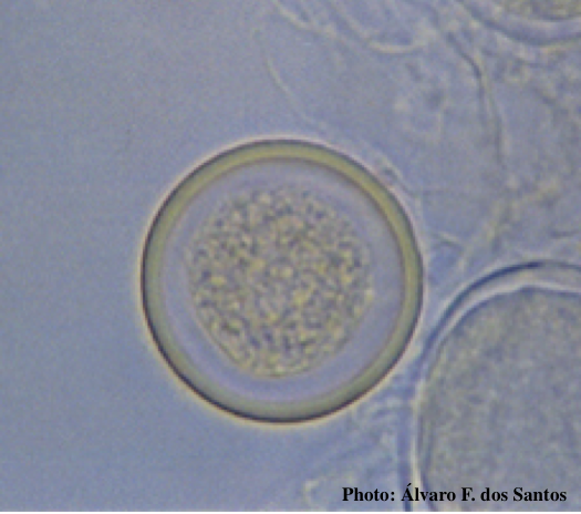 Globose chlamydospore (Fitopatol. bras. 2005) |
P. cambivora sporangium 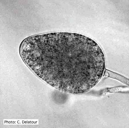 Ovoid non- papillate sporangia |
P. tentaculata sporangium  Papillate sporangium of P. tentaculata |