

Sporangia of P. lateralis showing internal and external proliferation
Photo Gallery
|
P. lateralis sporangia 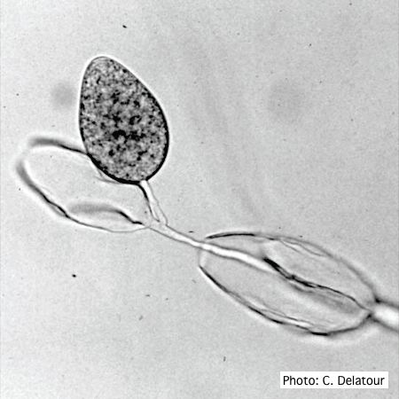 |
P. austrocedrae irregular sporangium 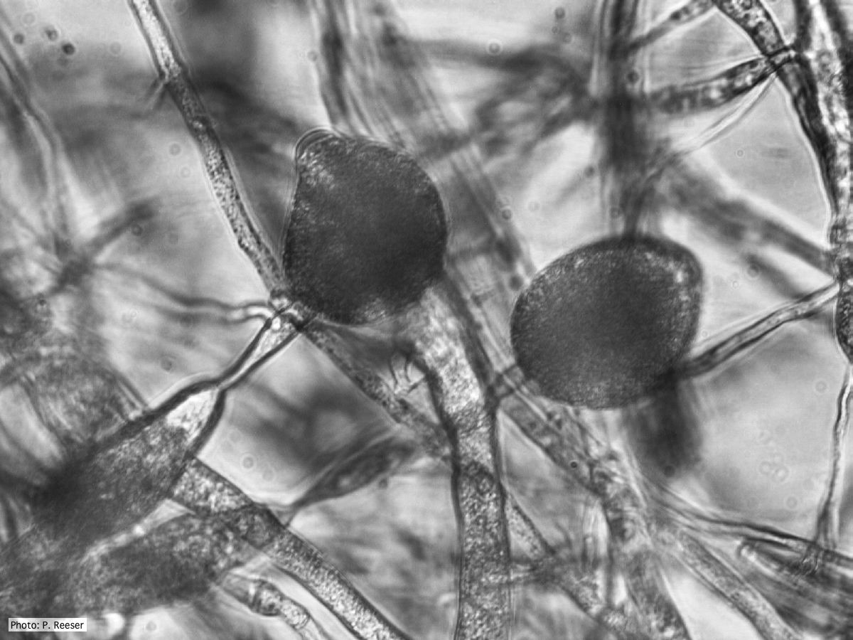 P. austrocedrae - irregular sporangium with lateral attachment and swelling in sporangiophore |
P. austrocedrae - Mal del ciprés in Argentina 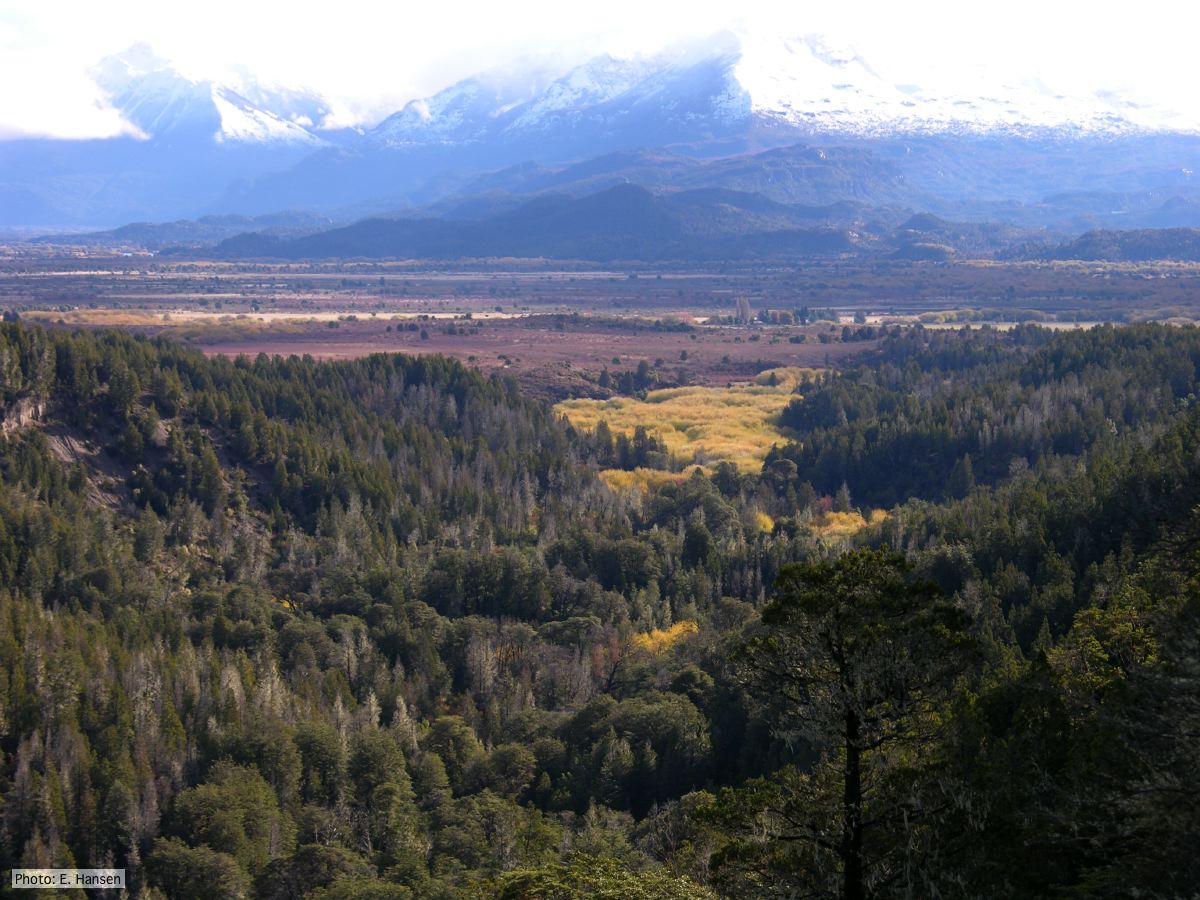 Mal del ciprés looking toward Rio Grande, Chubut Province, Argentina |
|
P. cactorum bleeding canker  Bleeding canker on European beech (Fagus sylvatica) |
P. megasperma sporangia 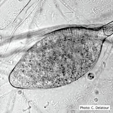 Ovoid, non-papillate sporangia showing internal proliferation of sporangiophore |
P. pluvialis sporangium 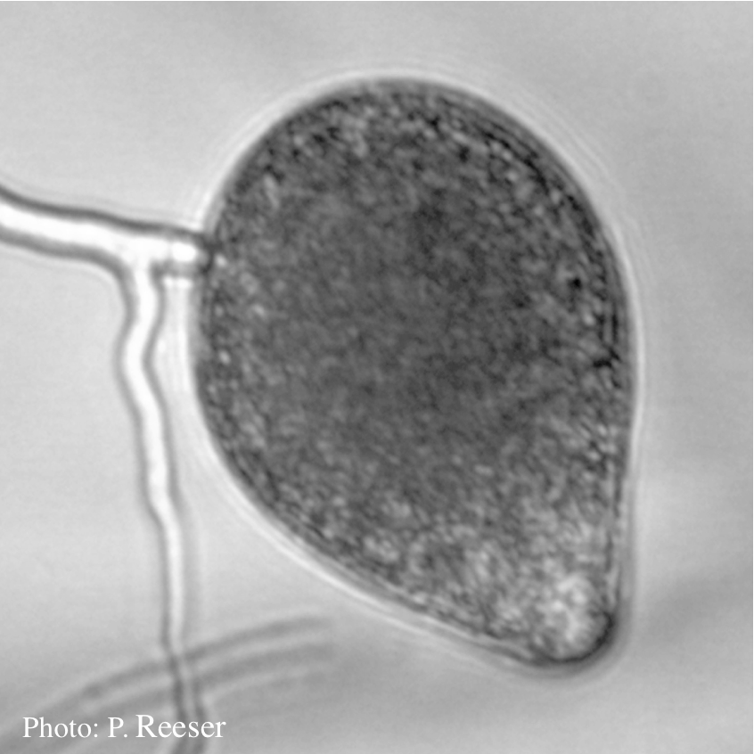 Sporangia showing typical ovoid shape and semi-papillate condition |
|
P. pinifolia sporangia 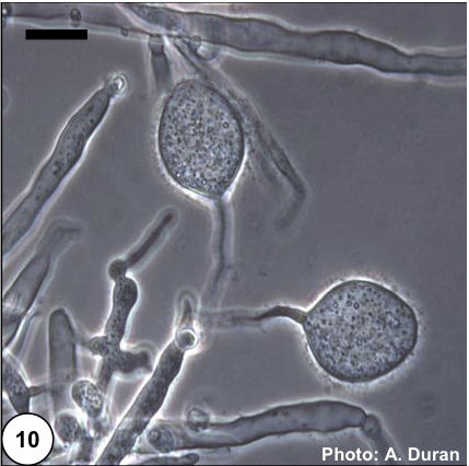 Non- papillate and caducous sporangia of Phytophthora pinifolia isolated from the infected P. radiata needles. |
P. nicotianae colony morphology on PDA 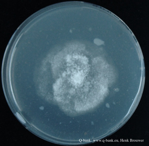 Phytophthora nicotianae CBS 321.49 PDA after 7 days at 24 degrees. Photo from Q-bank: www.q-bank.eu, Henk Brouwer (CBS-KNAW, Utrecht, The Netherlands) |
P. pinifolia sporangia 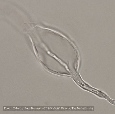 Sporangium with internal proliferation, photo from Q-bank, used with permission. |
|
P. cambivora active lesion on chinquapin  P. cambivora active lesion on chinquapin |
P. cambivora sporangium 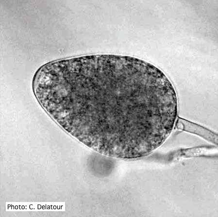 Ovoid non- papillate sporangia |
P. austrocedrae colony morphology on CMA 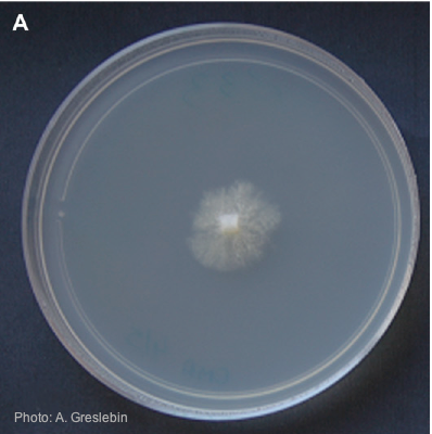 Colony morphology of P. austrocedrae at 16ºC after 4 weeks on CMA |