

Pinus radiata dead needles caused by DFP with healthy new growth from DFP
Photo Gallery
|
P. pinifolia on Pinus radiata  |
P. megakarya sporangia (photo from Q-bank, used with permission). 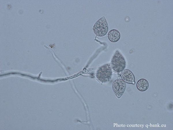 P. megakarya sporangia |
P. pseudosyringae sporangia  Ovoid, semipapillate sporangia showing sympodial development of sporangiophore |
|
P. kernoviae sporangium 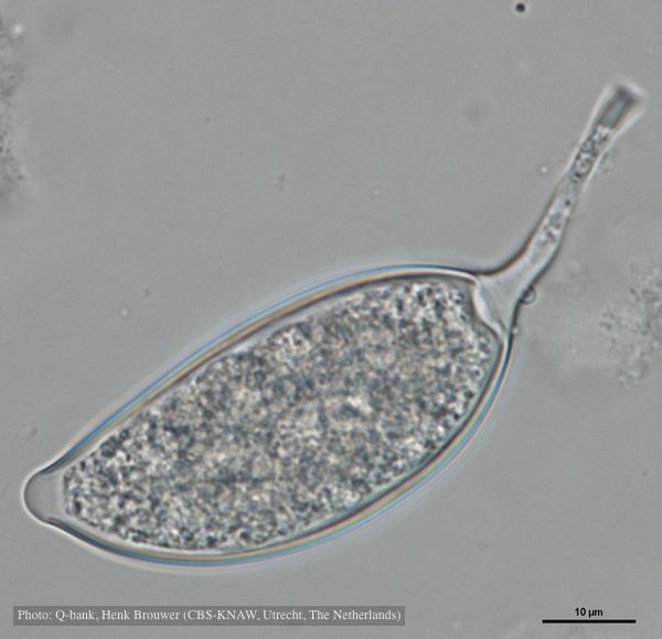 Asymmetrical sporangium, photo from Q-bank, used with permission |
P. agathidicida oospores in planta  Oospores in the roots of kauri seedlings inoculated with P. agathidicida. The root has been cleared with potassium hydroxide and bleached with peroxide before being stained with trypan blue (scale bar =100 µm). |
Growth of P. arenaria on CA  Colony morphology of Phytophthora arenaria after 7 days at 20°C on carrot agar |
|
P. cinnamomi hyphal swelling 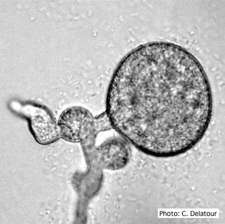 P. cinnamomi hyphal swelling (or thin walled chlamydospores) |
P. cambivora sporangium with sympodial proliferation 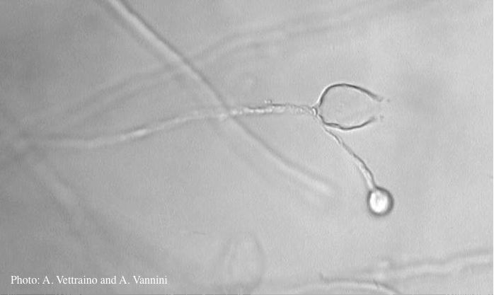 Empty sporagium showing sympodial proliferation |
P. pinifolia on Pinus radiata  Pinus radiata, note Stem canker associated with necrotic needles. |
|
P. austrocedrae - sporangia 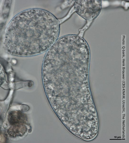 Sporangium with distorted shape, photo from Q-bank, used with permission. |
P. pinifolia on Pinus radiata 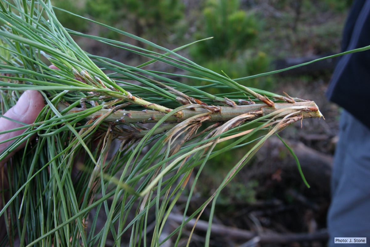 Pinus radiata, note grey and collapsed needle bases |
P. cambivora sporangium with internal extended proliferation 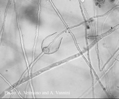 Empty sporangia showing internal extended proliferation |