

Sporangia showing typical ovoid shape and semi-papillate condition
Photo Gallery
Site will be retired 9/1/2026
This site is no longer being developed and will be retired on September 1, 2026. Please contact us if you have any questions or would like to provide support to continue the project.
|
P. pluvialis sporangium 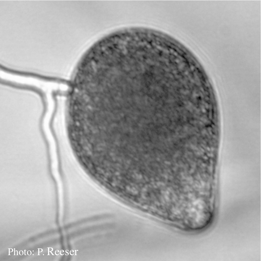 |
P. pluvialis symptoms on Douglas-fir 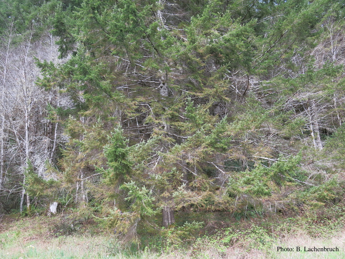 Red needle cast symptoms on Douglas-fir in western Oregon, 2015 |
P. pluvialis sporangia.  P. pluvialis sporangia on tape peel from infected Douglas-fir needle. |
|
P. pluvialis oogonium and antheridium 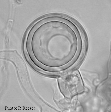 Oogonium and oospore with amphigynous antheridium |
P. pluvialis on Pinus radiata 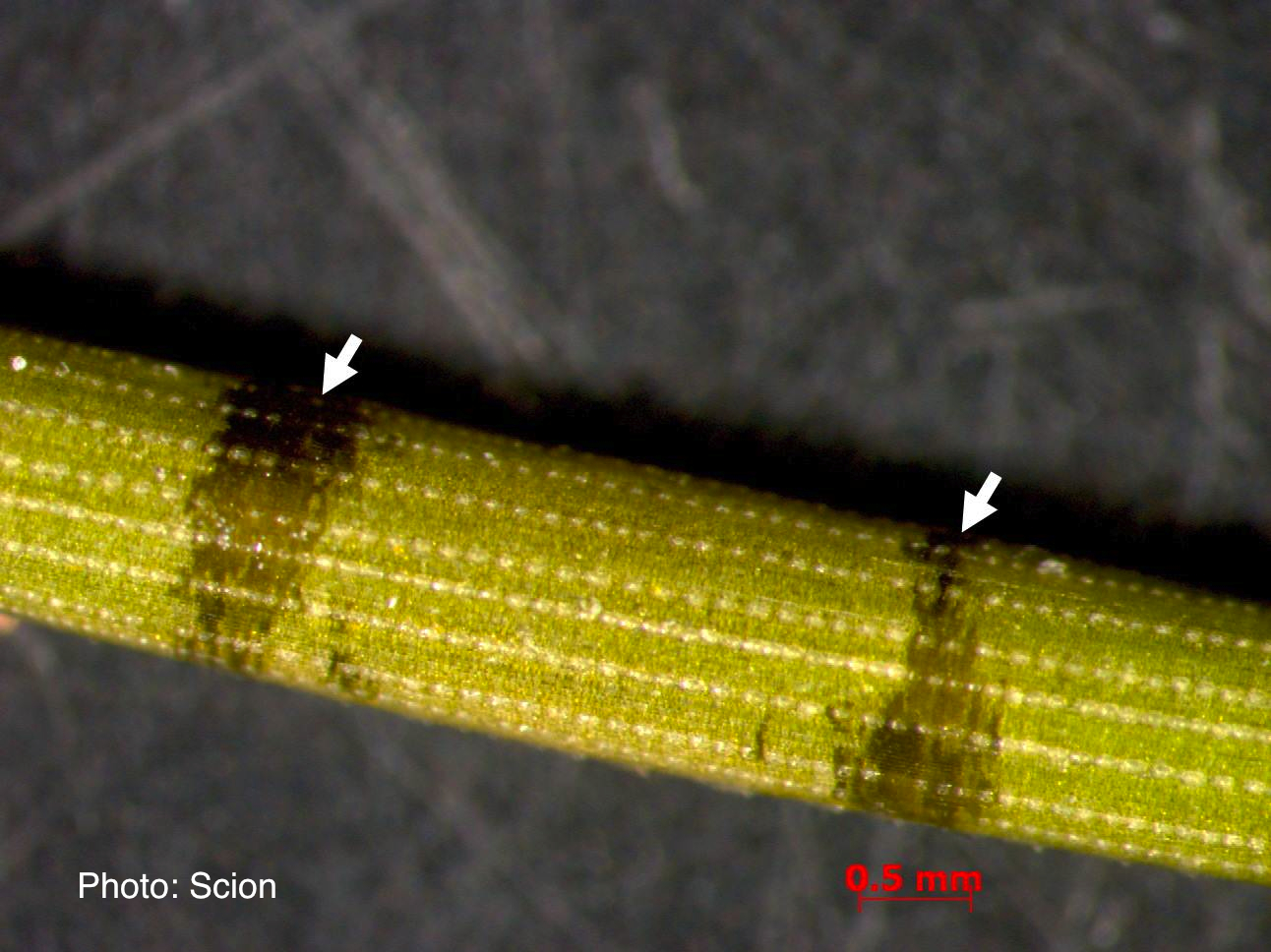 A Pinus radiata needle showing black resinous bands or marks consistent with the presence of red needle cast disease. |
P. pluvialis on Pinus radiata in New Zealand 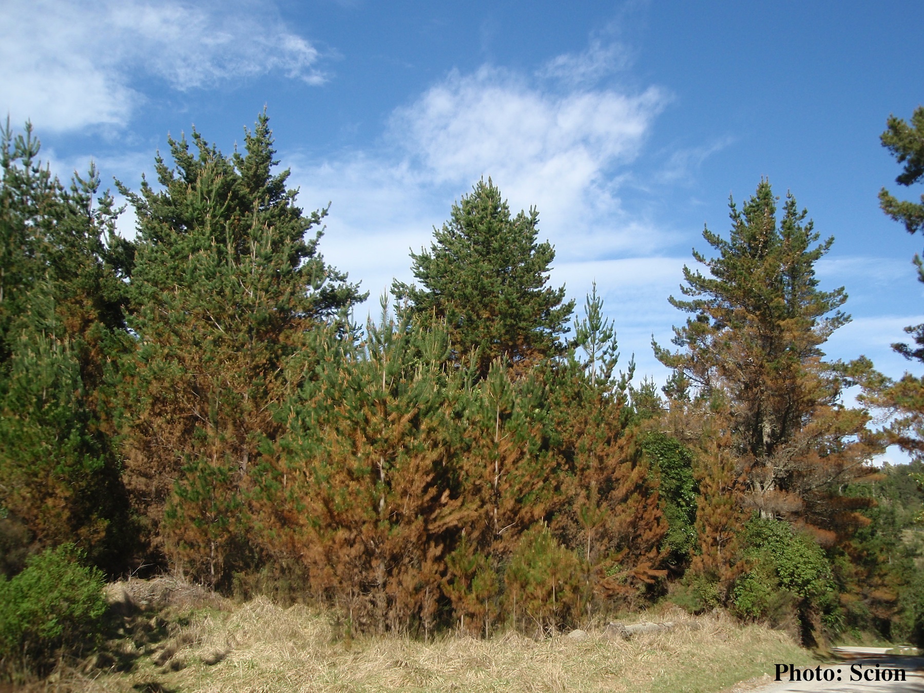 A stand of Pinus radiata trees affected by red needle cast disease. Note the reddish appearance of affected trees prior to needle drop. |
|
P. pluvialis on Pinus radiata in New Zealand 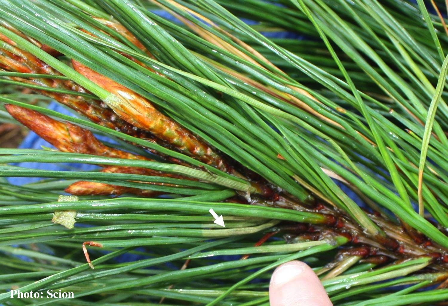 Lesions consistent with the presence of red needle cast disease are more abundant at the base of Pinus radiata needles as indicated by the arrow. |
P. pluvialis on Pinus radiata in New Zealand  Typical red needle cast symptoms along a twig. Lesions begin at the base of the needle which subsequently turns brown and is cast from the twig. |
P. pluvialis sporangium 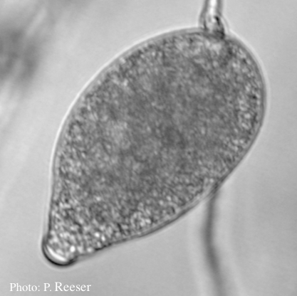 Sporangium showing typical ovoid shape and semi-papillate condition |
|
P. pluvialis hyphal swellings 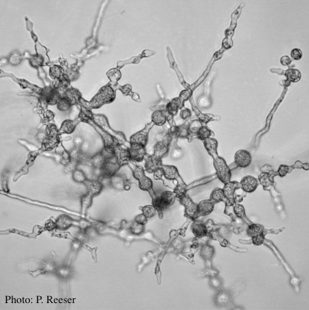 P. pluvialis hyphal swellings on agar |
P. pluvialis symptoms on Douglas-fir 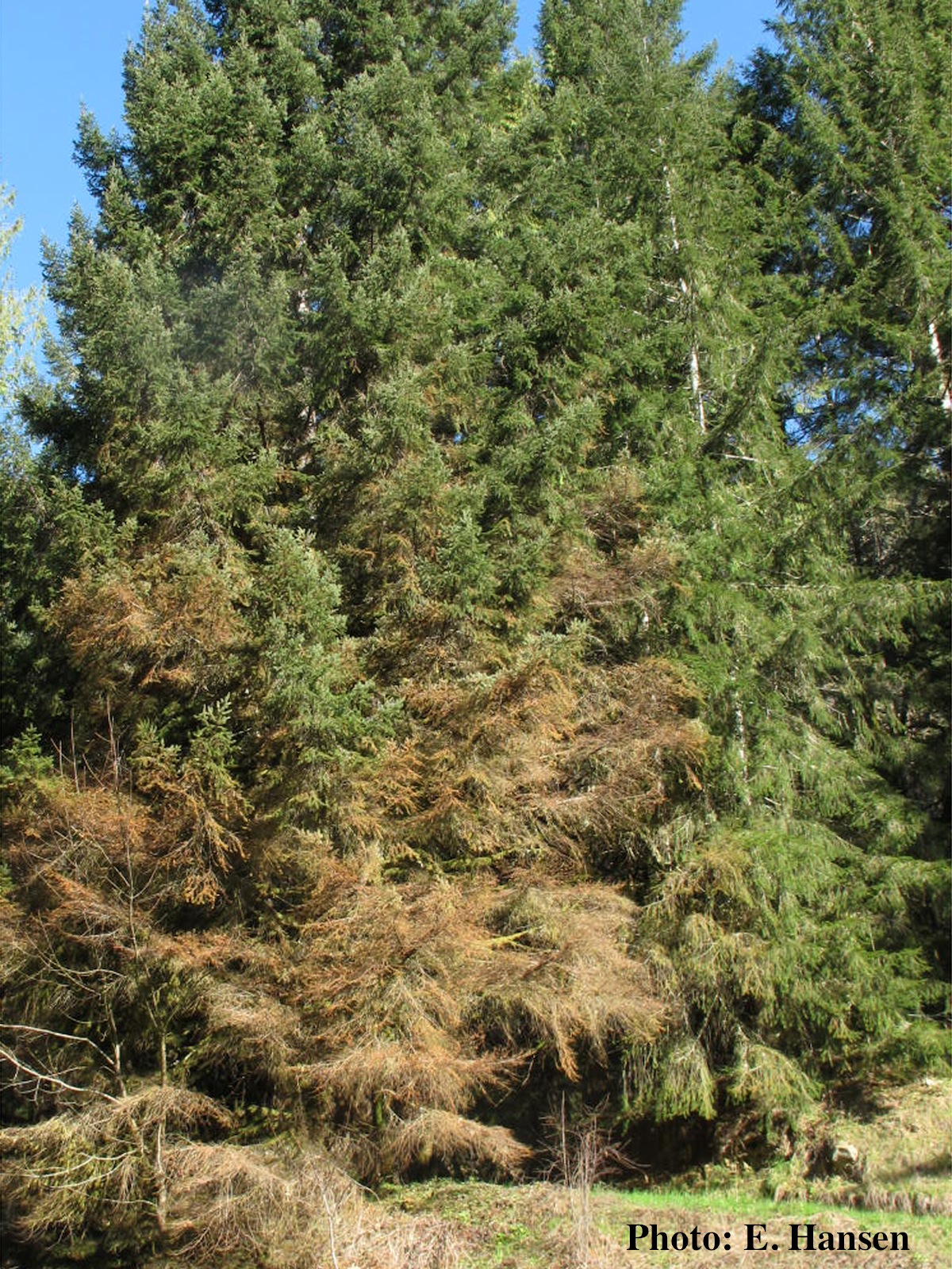 Red needle cast symptoms on Douglas-fir in western Oregon, 2015 |
P. pluvialis on Pinus radiata in New Zealand 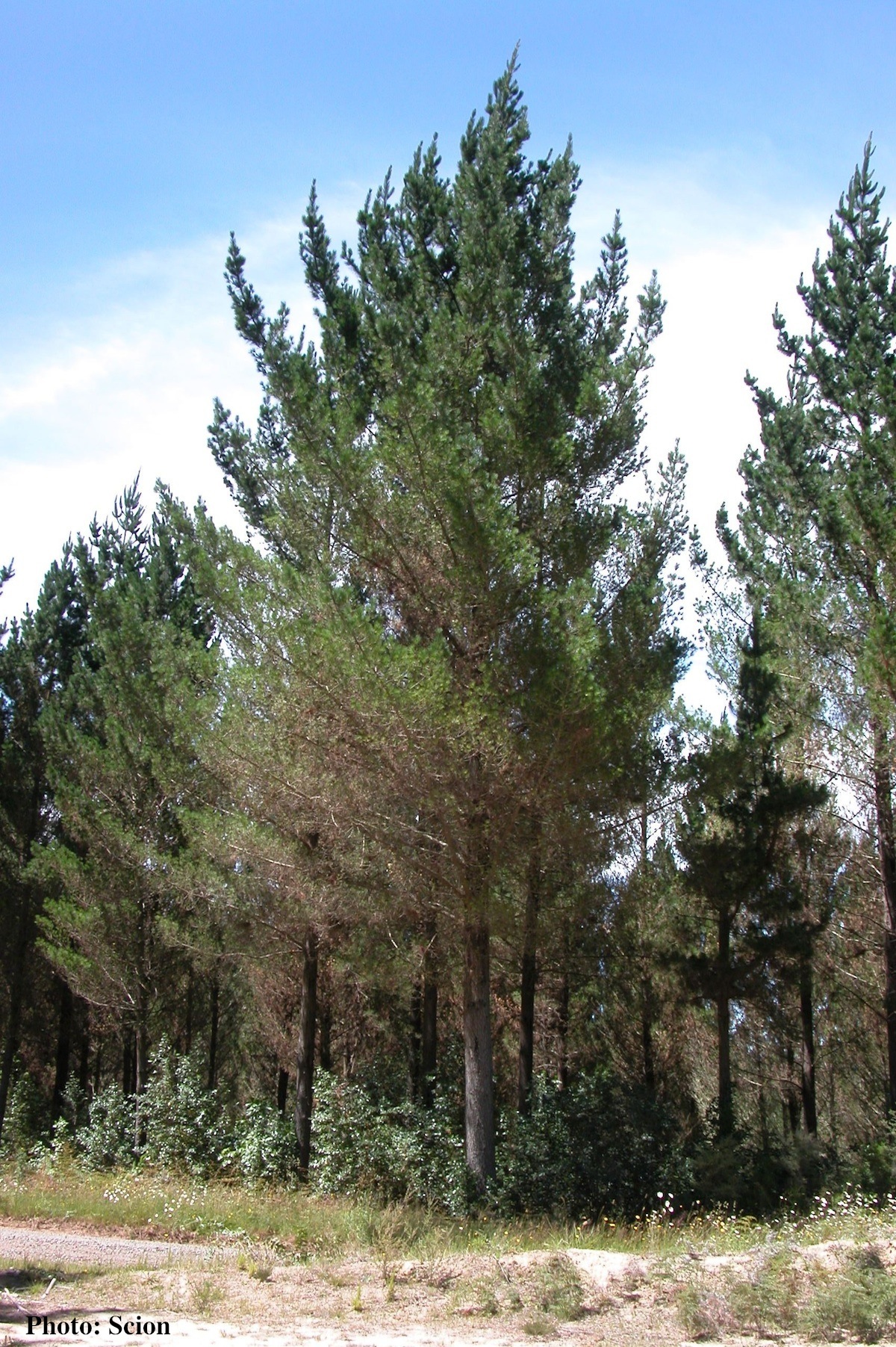 A stand of Pinus radiata trees affected by red needle cast disease. Note that frequently only the lower part of the crown is affected. |