

P. pseudotsugae colony growth on PDA agar
Photo Gallery
|
P. pseudotsugae colony morphology on PDA  |
P. cinnamomi hyphal swelling 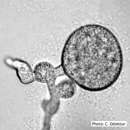 P. cinnamomi hyphal swelling (or thin walled chlamydospores) |
P. alni canker on gray alder 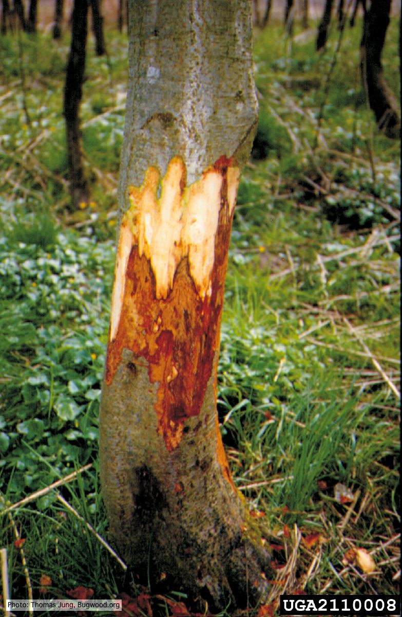 Grey alder (A. incana) with collar rot caused by P. alni |
|
P. cactorum sporangia 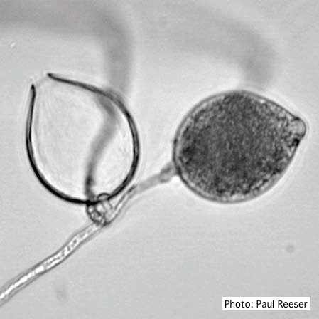 Broadly ovoid, papillate sporangia in water. |
P. cryptogea sporangia  Sporangiophore showing internal proliferation through empty sporangia after zoospore release |
P. kernoviae disease on European beech 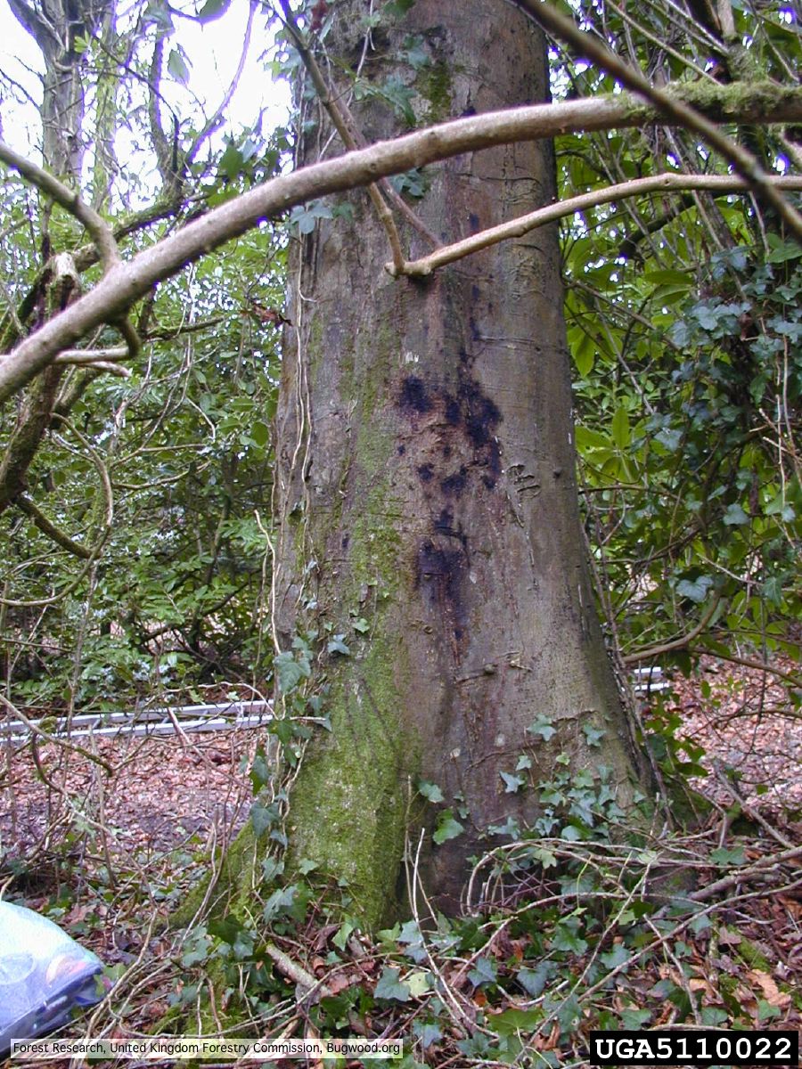 Bleeding lesion on trunk of Fagus sylvatica |
|
P. alni symptoms on European Alder  Mature, riparian common alder (A. glutinosa) stand heavily impacted by root and collar rot caused by P. alni |
P. cactorum oogonium 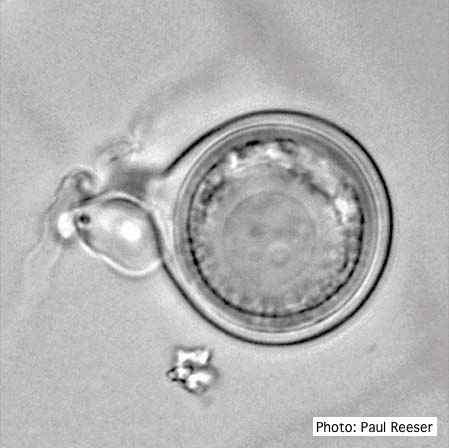 Oogonium with paragynous antheridia close to oogonial stalk. Oospores are slightly aplerotic. |
P. cambivora colony morphology on PDA 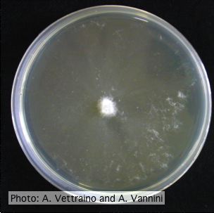 Appressed colony morphology at 14 days at 20°C on PDA |
|
P. tentaculata chlamydospore 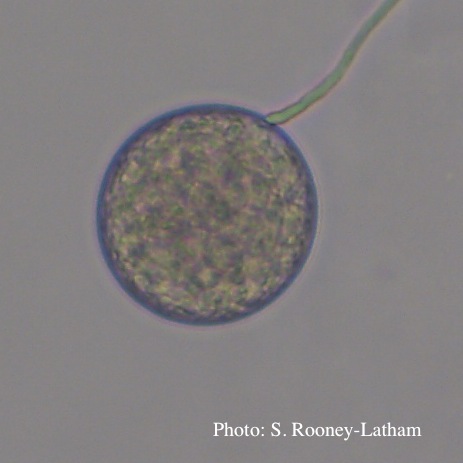 Terminal chlamydospore of P. tentaculata |
P. cryptogea hyphal swellings 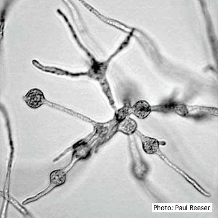 Cluster of small, angular to globose hyphal swellings formed in water |
P. pinifolia colony morphology on PDA 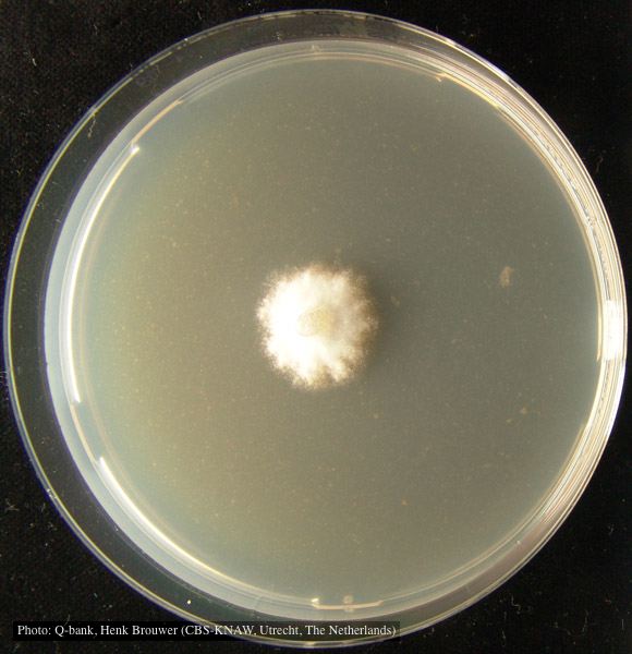 Colony pattern after 7 days on PDA at 24C, photo from Q-bank, used with permission. |