

Papillate, non-caducous sporangium with differentiated content; photo used with permission from Q-bank
Photo Gallery
|
P. katsurae sporangia 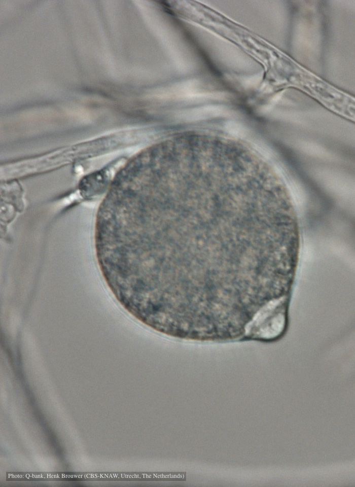 |
P. agathidicia sporangia 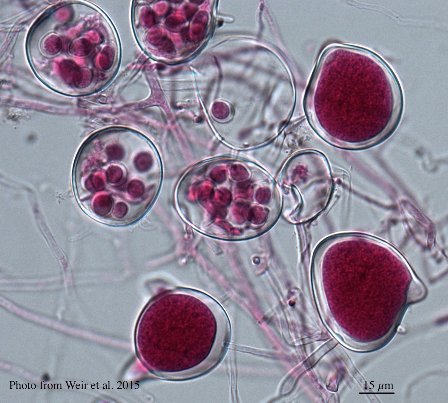 Differentiation of the cytoplasm within papillate sporangia into acid fuchsin stained zoospores |
P. cactorum oogonium 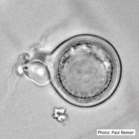 Oogonium with paragynous antheridia close to oogonial stalk. Oospores are slightly aplerotic. |
|
P. austrocedrae colony morphology on PDA  Colony morphology of P. austrocedrae at 16 C after four weeks on PDA |
P. kernoviae disease on European beech 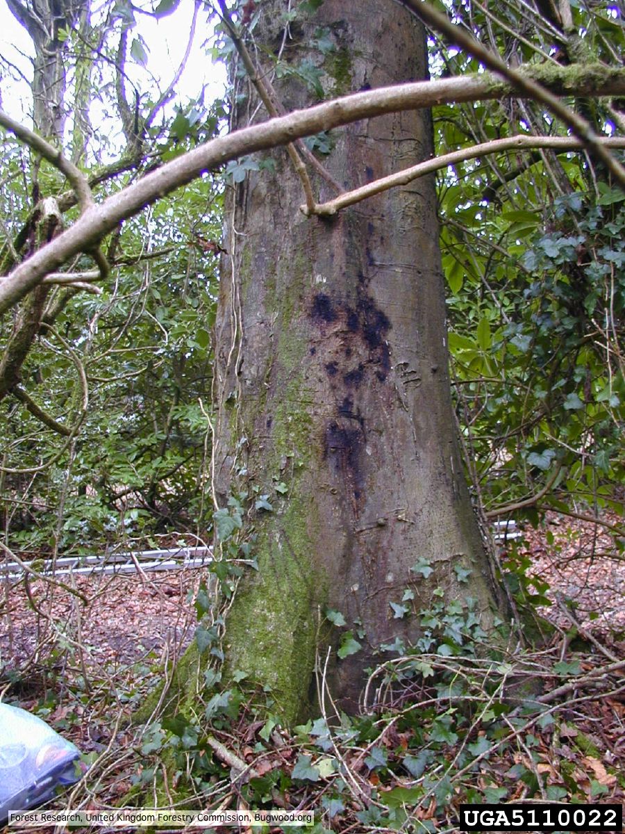 Bleeding lesion on trunk of Fagus sylvatica |
P. alni symptoms on European Alder 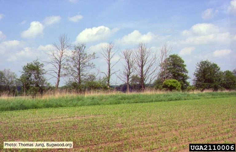 Mature, riparian common alder (A. glutinosa) stand with high impact of Phytophthora root and collar rot. |
|
P. kernoviae leaf wilt 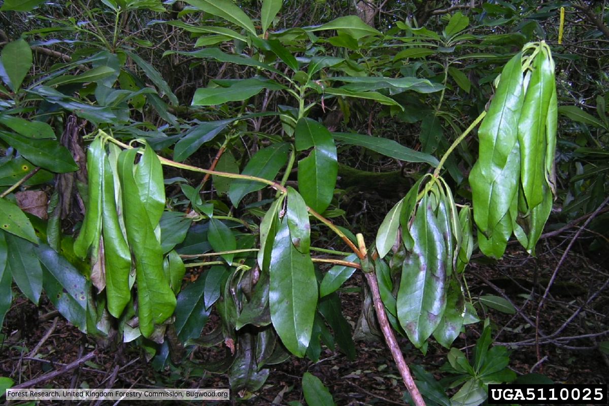 Wilted leaf of infected rhododendron |
P. megakarya disease symptoms on Theobroma cacao

Symptoms of black pod disease of cocoa (T. cacao)
|
P. cambivora sporangium 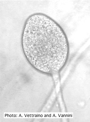 Ovoid non-papillate sporangia with well-rounded base and simple sporangiophore |
|
P. lateralis on Port Orford cedar 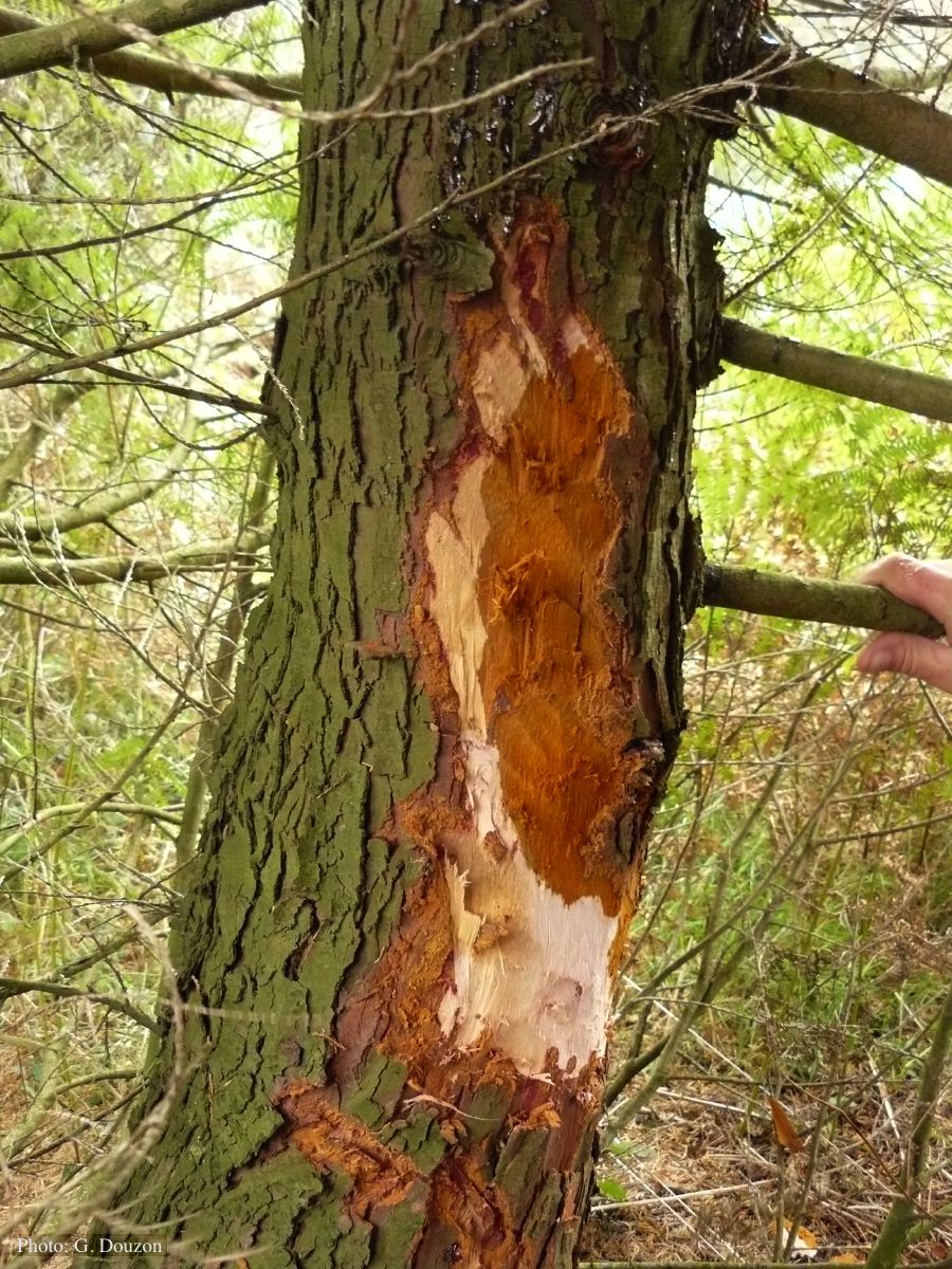 Bole lesion on Chaemacyparis lawsoniana in Lopérec, France |
P. pluvialis colony morphology on carrot agar  Colony morphology on carrot agar at 20 days |
P. cactorum bleeding canker 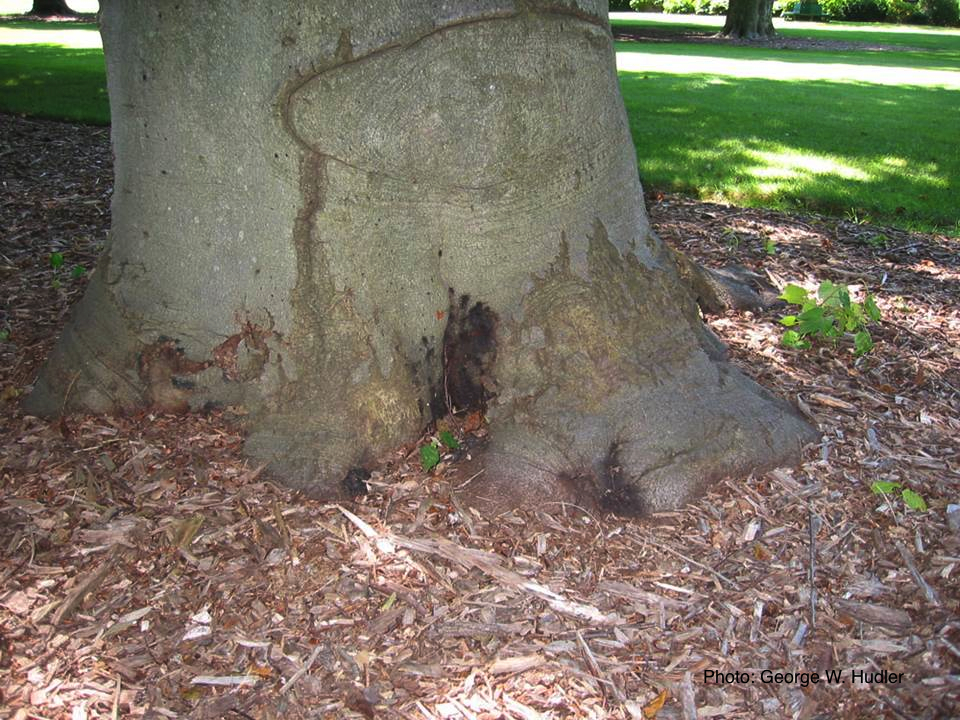 Bleeding canker on European beech (Fagus sylvatica) |