

P. austrocedrae. Morphology of oogonia, oospores and antheridia. Bar: 10 mm. Greslebin et al. 2007
Photo Gallery
|
P. austrocedrae oogonia drawing  |
P. cryptogea hyphal swellings 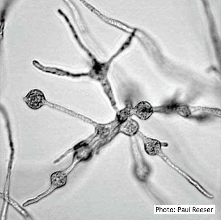 Cluster of small, angular to globose hyphal swellings formed in water |
P. cactorum bleeding canker 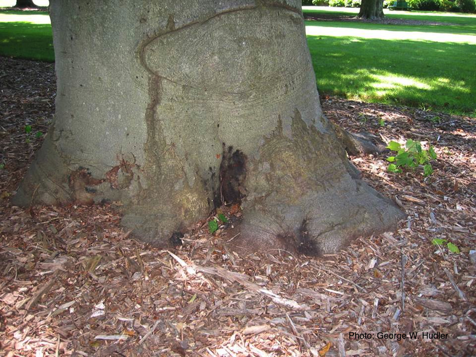 Bleeding canker on European beech (Fagus sylvatica) |
|
P. cambivora disease symptoms 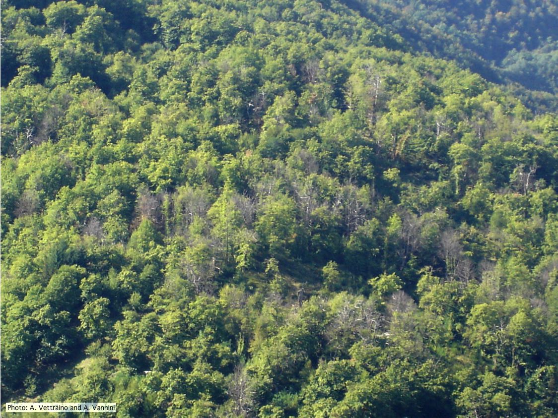 Ink disease impact in sweet chestnut forest in Italy |
P. ramorum colony morphology on V8 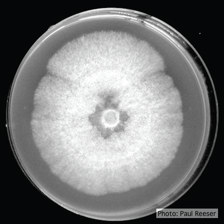 P. ramorum colony morphology on V8 |
P. austrocedrae semipapillate sporangium  P. austrocedrae - semipapillate sporangium with off-center attachment. |
|
P. kernoviae leaf wilt 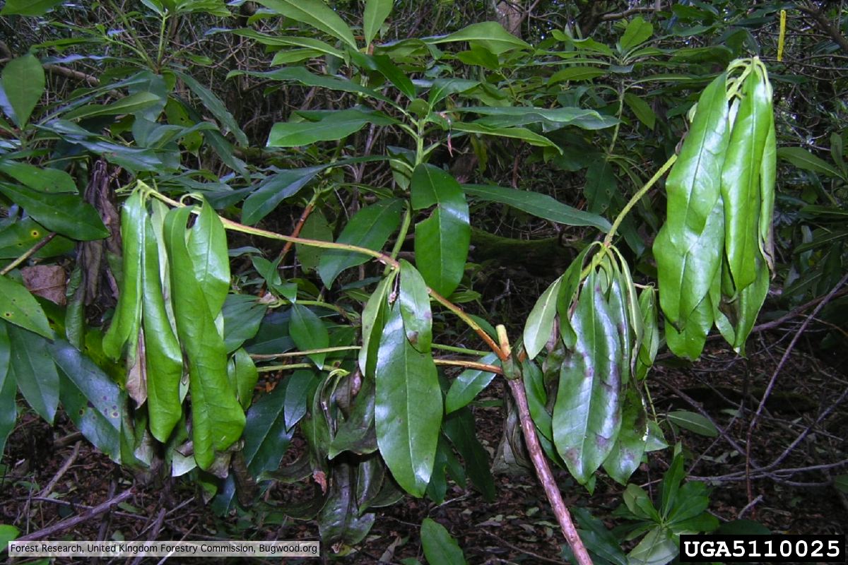 Wilted leaf of infected rhododendron |
Mal del ciprés, dead and dying trees along river 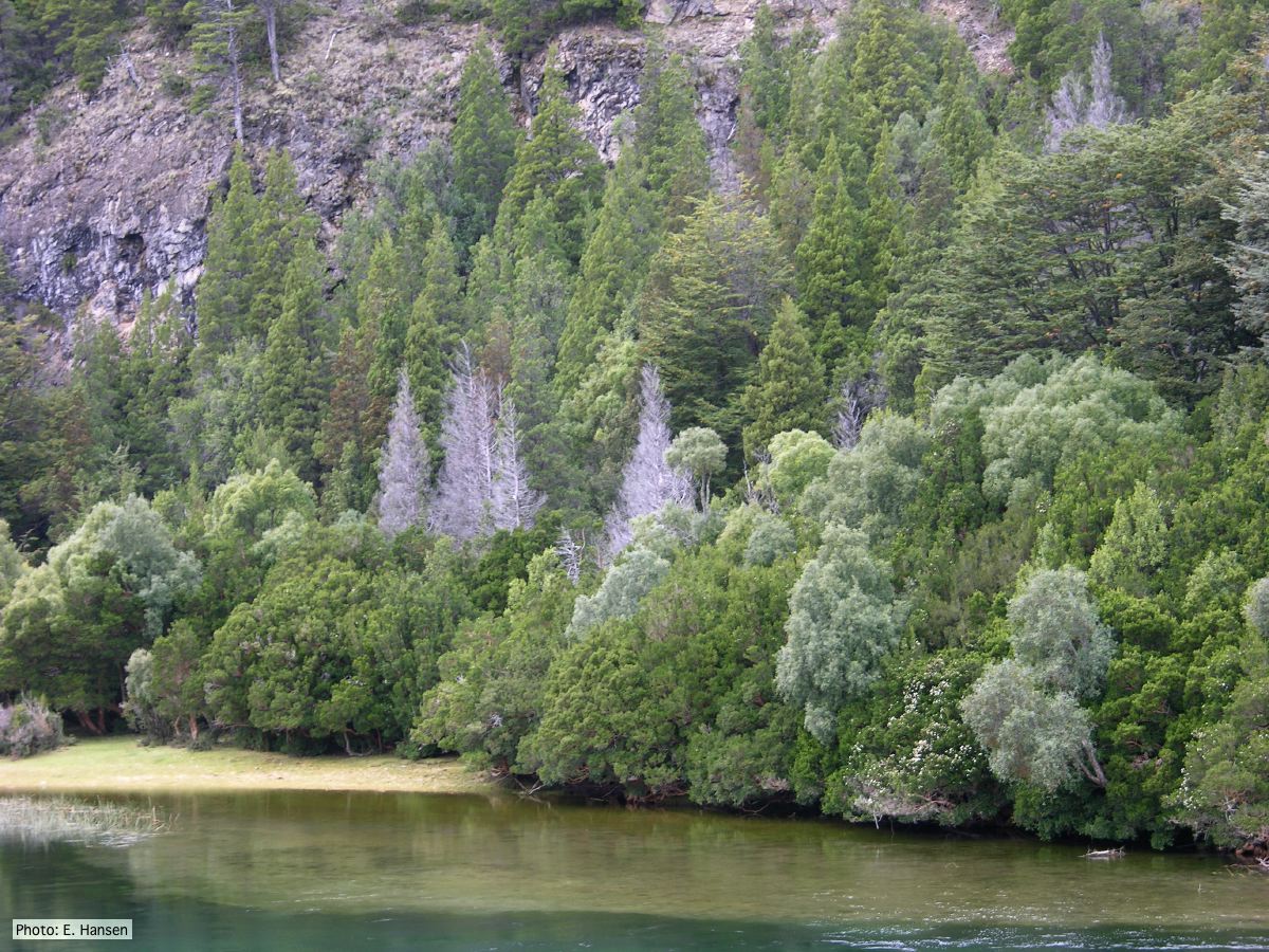 Mal del ciprés, dead and dying trees along river |
P. cambivora older hyphae  Old septate hyphae |
|
P. katsurae disease symptoms  Infected chestnut tree with girdling canker on stem |
P. cactorum colony morphology on PDA 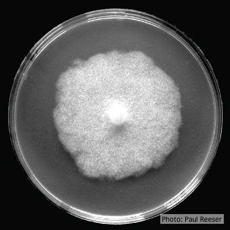 Colony morphology on PDA at 14 days |
Comparative gametangial morphology of Phytophthora Clade 5 species  Comparative gametangial morphology of Phytophthora Clade 5 species, with SEM (top) and light microscopy (bottom). P. heveae has smooth walled oogonia with funnel-shaped, amphigynous antheridia. P. agathidicida has mildly stipulate oogonia with globose amphigynous antheridia. P.cocois has mildly bullate oogonia with reflexed amphigynous antheridia. P. castaneae has coarsely bullate oogonium with rugose protuberances and narrow amphigynous antheridia (Weir et al. 2015). |