

Differentiation of the cytoplasm within papillate sporangia into acid fuchsin stained zoospores
Photo Gallery
|
P. agathidicia sporangia 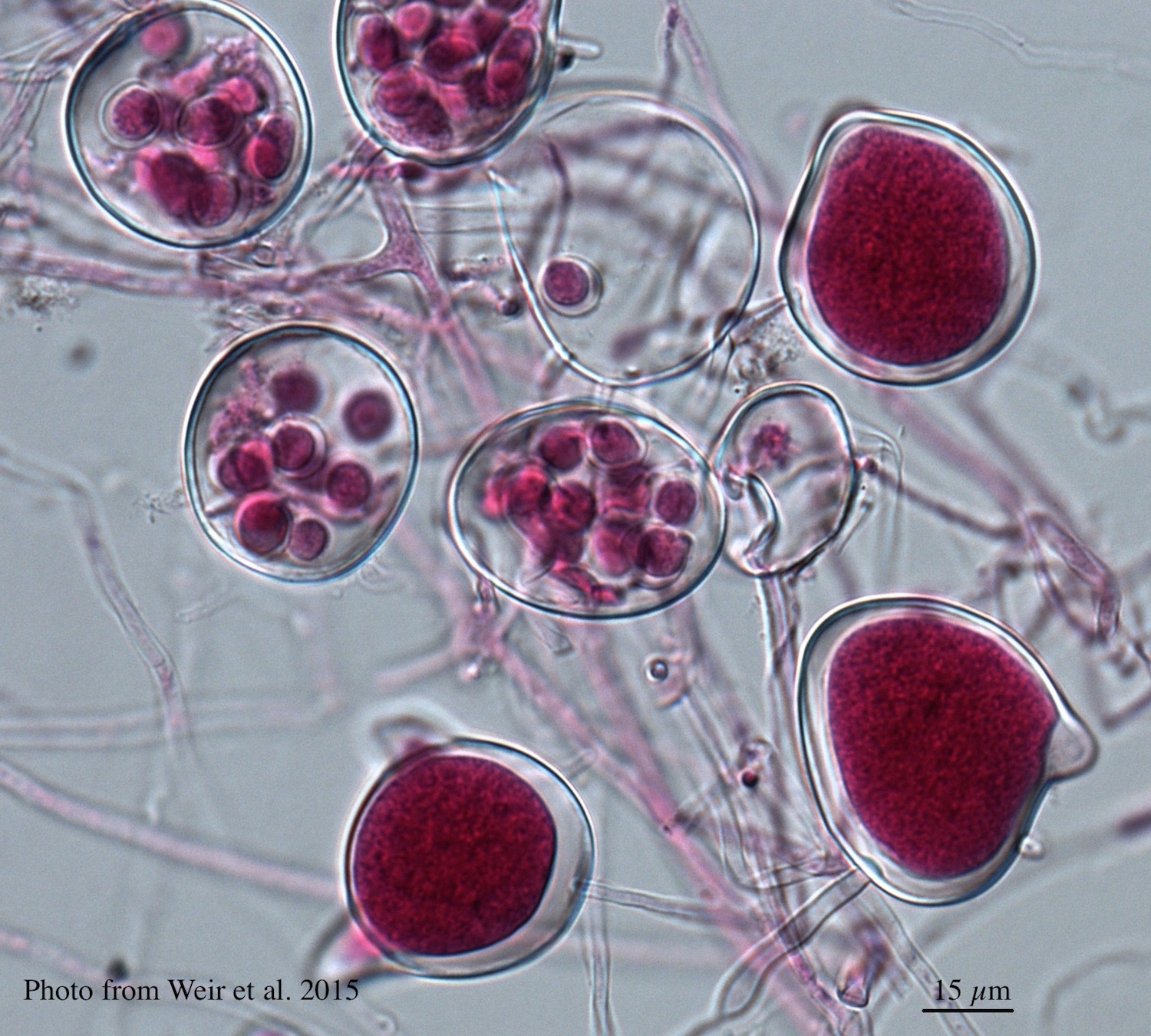 |
P. pluvialis on Pinus radiata in New Zealand  Pinus radiata needles showing colour changes following infection with red needle cast disease. The tissues around the initial infection at the base or along the needle senesce, and change yellow and then brown as indicated by the arrows before the needles cast. |
P. pseudotsugae paragynous oogonium 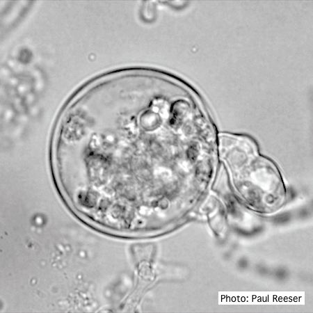 P. pseudotsugae oogonium with paragynous antheridia, note the disrupted contents of the abortive oogonia |
|
P. kernoviae colony morphology on V8 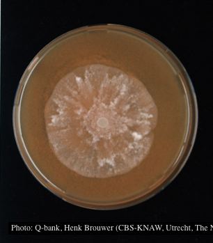 Colony morphology at 7 days at 18°C on V8, photo from Q-bank, used with permission. |
Phytophthora taxon Agathis bole canker  Canker on a Kauri tree, New Zealand |
P. agathidicia oogonia 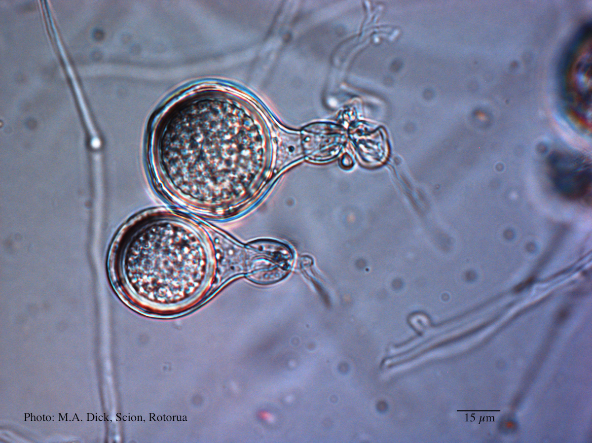 Light micrograph of P. agathidicida oospore (Scale bar equals 15 µm) |
|
P. siskiyouensis sporangium  P. siskiyouensis sporangium with lateral semi-papilla and subterminal, sub-basal insertion in the sporangiophore |
P. pinifolia on Pinus radiata 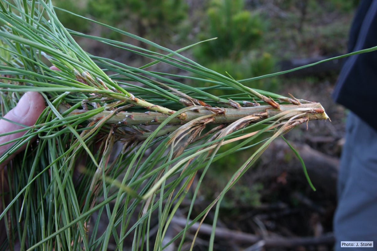 Pinus radiata, note grey and collapsed needle bases |
P. nicotianae colony morphology on V8 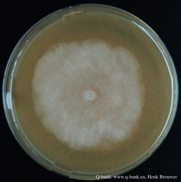 Phytophthora nicotianae CBS 321.49 V8 after 7 days at 24 degrees. Photo from Q-bank: www.q-bank.eu, Henk Brouwer (CBS-KNAW, Utrecht, The Netherlands) |
|
P. austrocedrae - Mal del ciprés, stages of decline 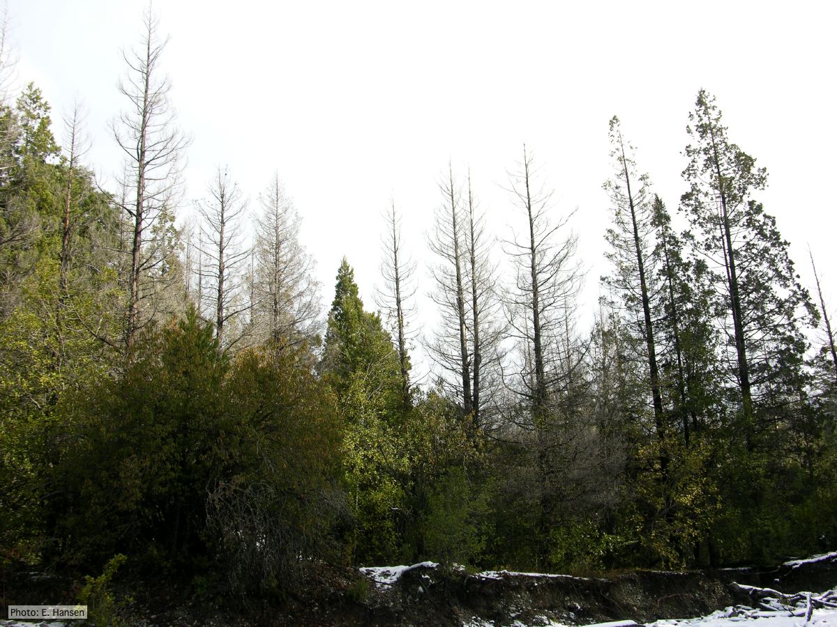 Colony morphology of P. austrocedrae at 16 C after four weeks on PDA |
P. kernoviae leaf wilt 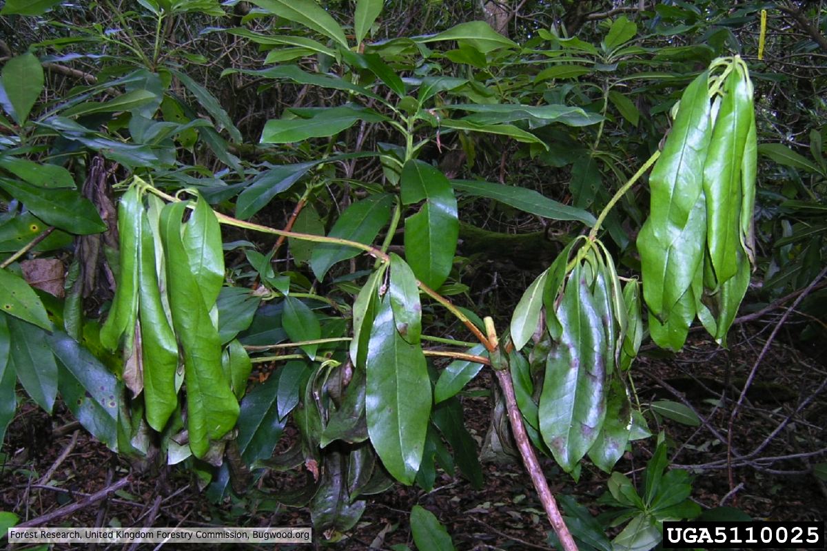 Wilted leaf of infected rhododendron |
P. agathidicida oospores  Oospores of P. agathidicida in the roots of kauri seedlings inoculated with P. agathidicida. The root has been cleared with potassium hydroxide and bleached with peroxide, before being stained with Trypan Blue |