

P. palmivora colony morphology on PDA
Photo Gallery
|
P. palmivora colony morphology on PDA 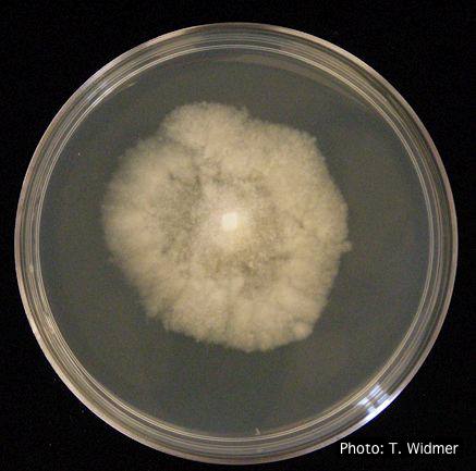 |
Phytophthora chlamydospora sporangium 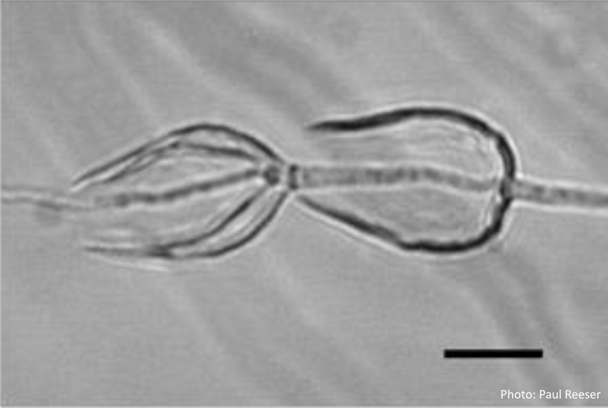 Phytophthora chlamydospora sporangia in water, showing internal proliferation. Bar is 20 µm. |
P. cinnamomi hyphal swellings 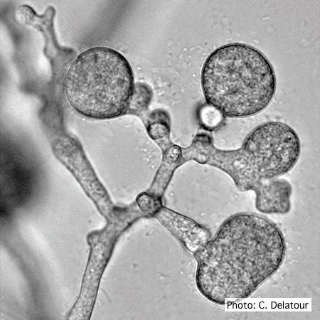 P. cinnamomi hyphal swellings (or thin walled chlamydospores) |
|
P. nicotianae oogonia 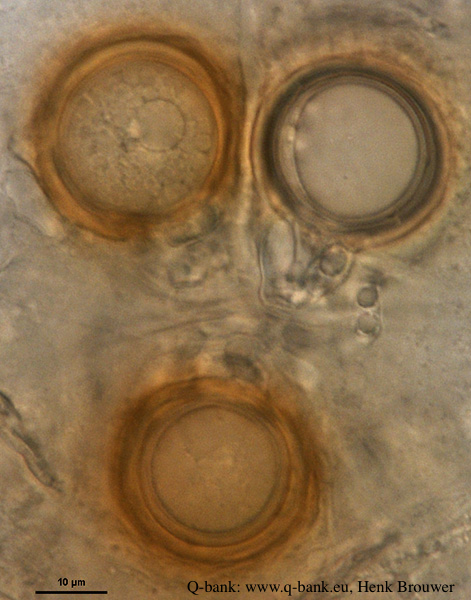 P. nicotianae oogonia 100x. Photo from Q-bank: www.q-bank.eu, Henk Brouwer (CBS-KNAW, Utrecht, The Netherlands) |
P. lateralis on Port Orford cedar  Localized branch infection of Chaemacyparis lawsoniana in Lopérec, France |
P. frigida symptoms 3 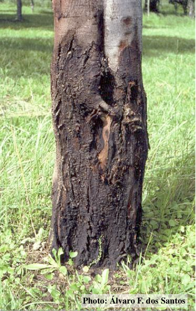 Black wattle bark with symptoms of gummosis |
|
P. austrocedrae colony morphology on Tomato juice agar with B sitosterol 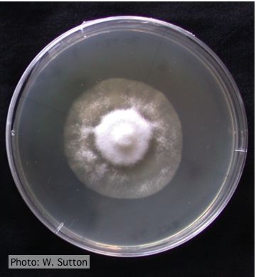 Colony morphology of P. austrocedrae at 16 ºC after 4 weeks on Tomato juice agar with B sitosterol |
P. cambivora oogonium 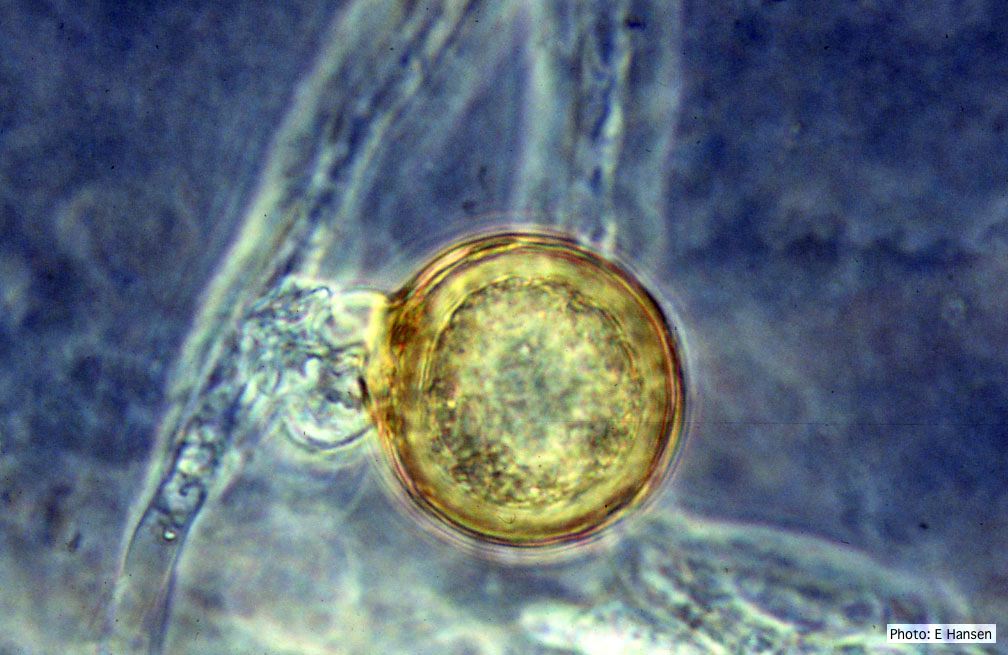 P. cambivora oogonium with antheridium |
Dying Port Orford Cedar trees 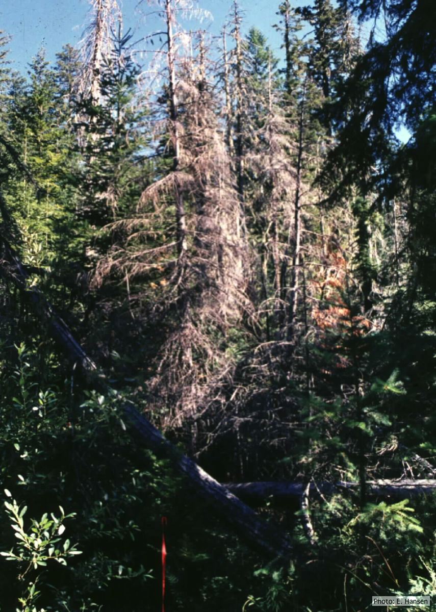 Dead Chamaecyparis lawsoniana trees |
|
P. palmivora chlamydospore 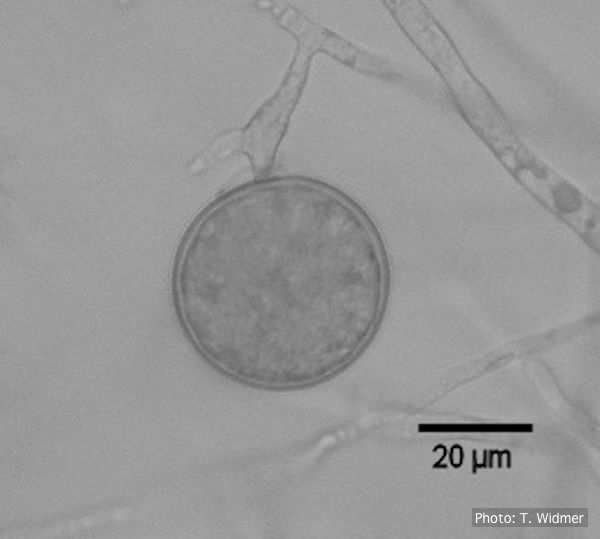 Terminal chlamydospore of P. palmivora |
P. ramorum canker 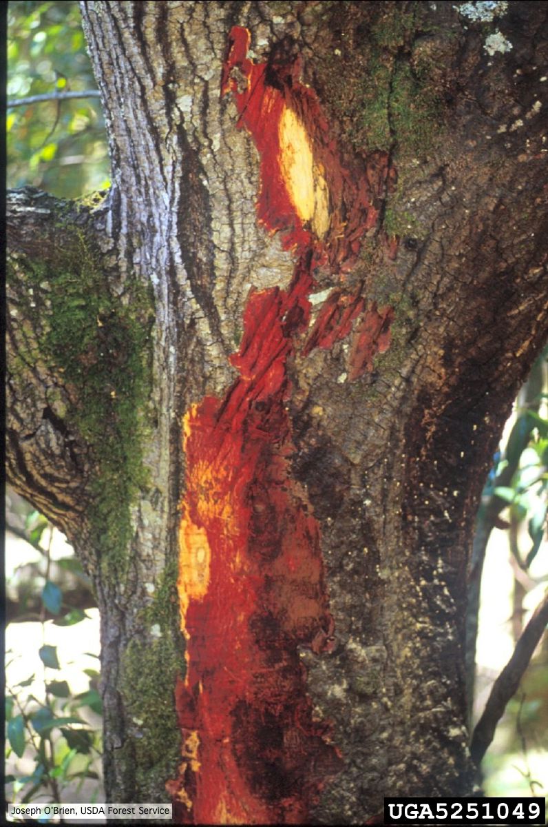 Bark discoloration and zone lines in coast live oak (Quercus agrifolia) |
P. pluvialis symptoms on Douglas-fir needles 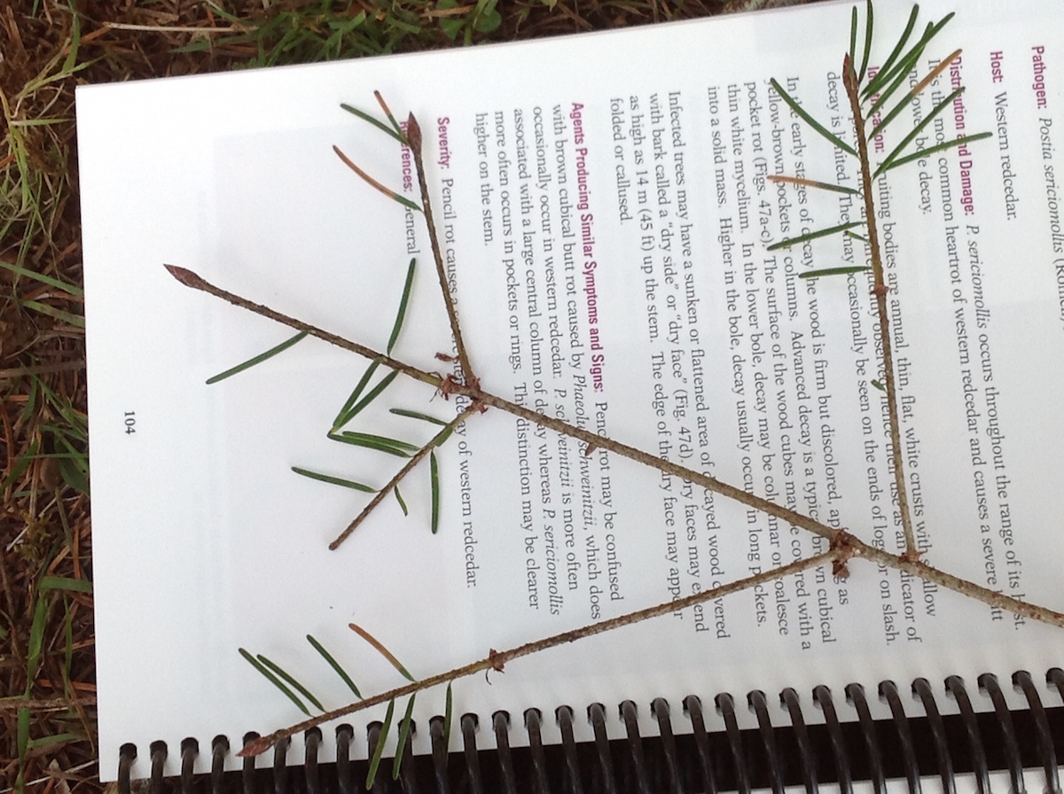 Symptoms of red needle cast on Douglas-fir needles |