

Colony morphology on PDA at 7 days
Photo Gallery
|
P. megasperma colony morphology on PDA  |
P. pluvialis symptoms on Douglas-fir  P. pluvialis symptoms of red needle cast on Douglas-fir, western Oregon 2015 |
P. pluvialis symptoms on Douglas-fir needles  Symptoms of red needle cast on Douglas-fir needles |
|
P. cambivora disease symptoms 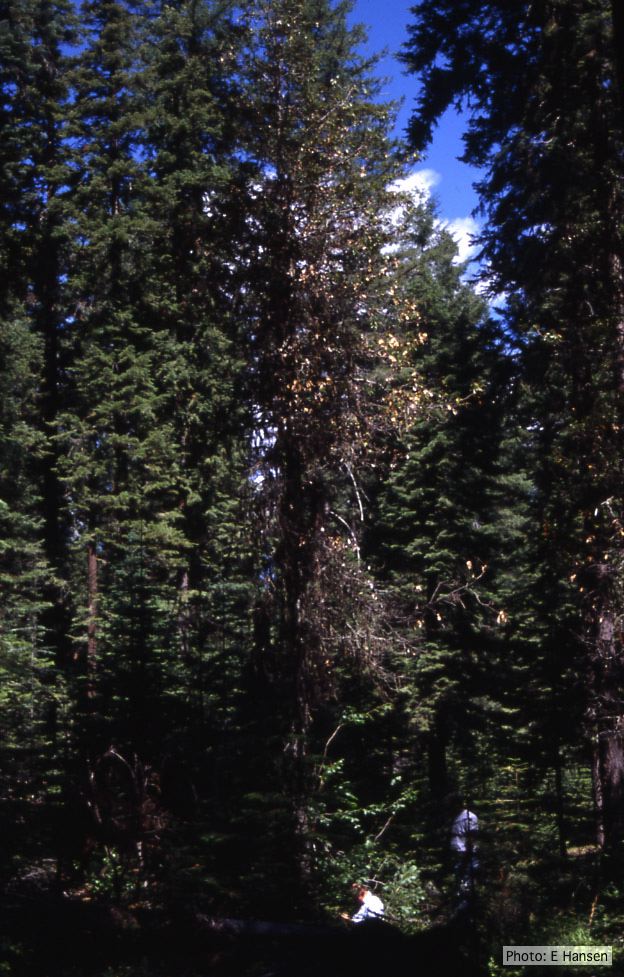 Dead and dying chinquapin infected with P. cambivora |
P. austrocedrae - sporangia drawings 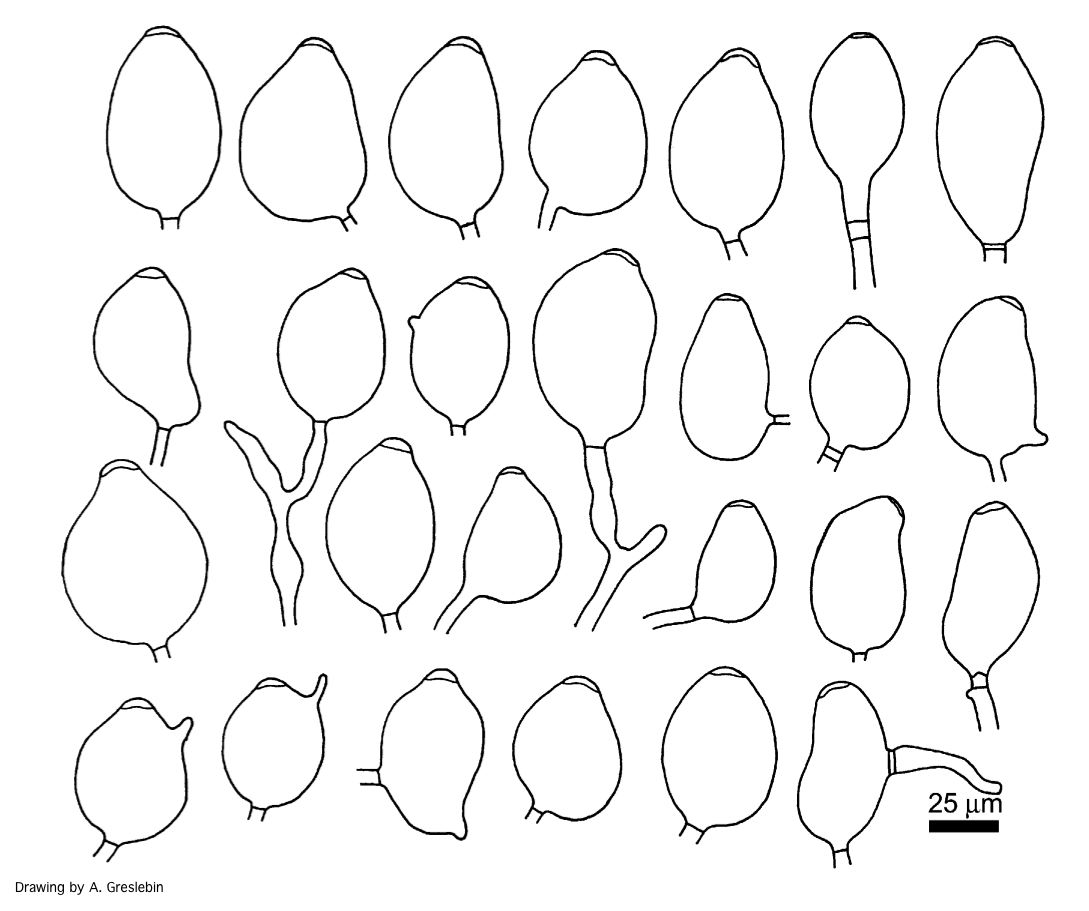 Phytophthora austrocedrae. Morphology of sporangia. Bar: 25 mm. Greslebin et al. 2007 |
P. chlamydospora colony morphology on carrot agar 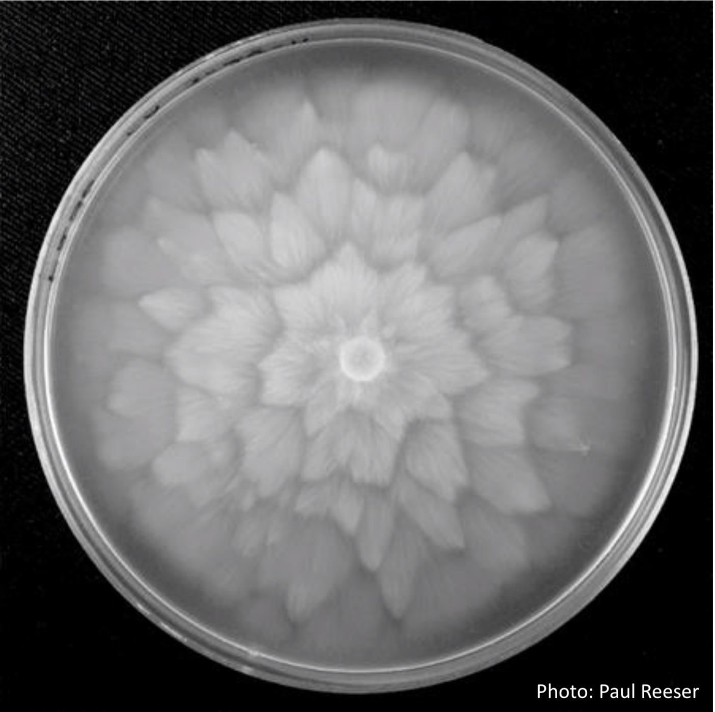 P. chlamydospora colony morphology on carrot agar |
|
P. lateralis on Port Orford cedar 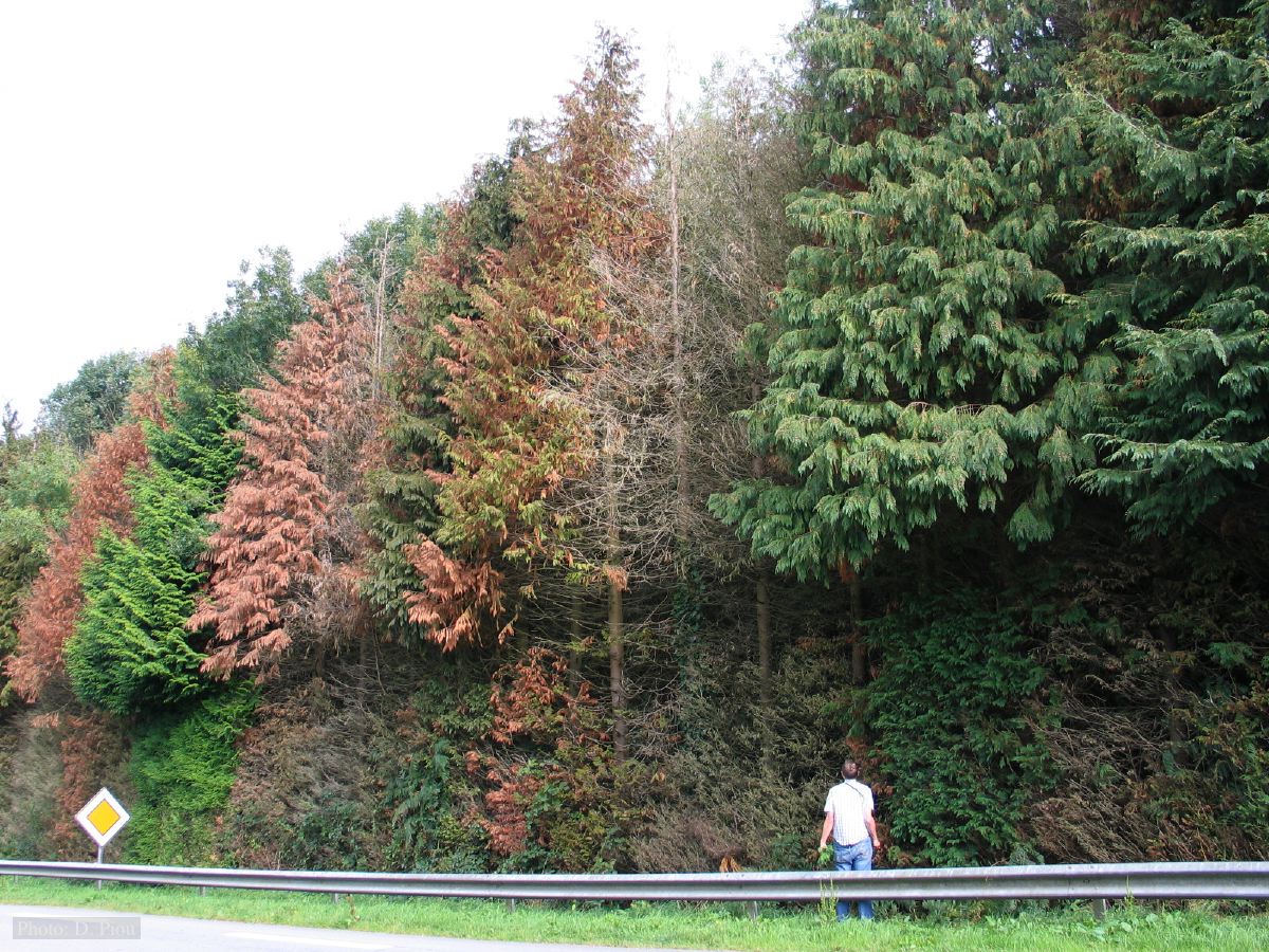 Typical decline of Chaemacyparis lawsoniana in Landrévarzec, France. |
P. megasperma sporangia 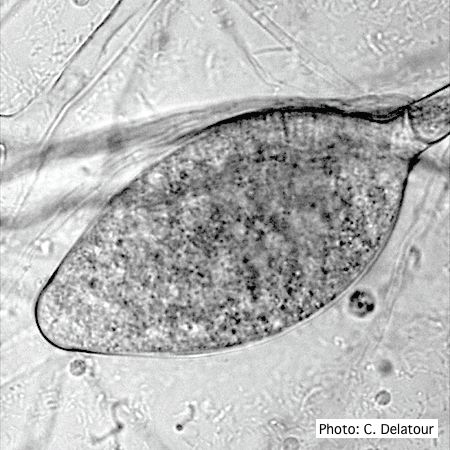 Ovoid, non-papillate sporangia showing internal proliferation of sporangiophore |
Comparative gametangial morphology of Phytophthora Clade 5 species  Comparative gametangial morphology of Phytophthora Clade 5 species, with SEM (top) and light microscopy (bottom). P. heveae has smooth walled oogonia with funnel-shaped, amphigynous antheridia. P. agathidicida has mildly stipulate oogonia with globose amphigynous antheridia. P.cocois has mildly bullate oogonia with reflexed amphigynous antheridia. P. castaneae has coarsely bullate oogonium with rugose protuberances and narrow amphigynous antheridia (Weir et al. 2015). |
|
P. tentaculata disease symptoms on sticky monkey flower 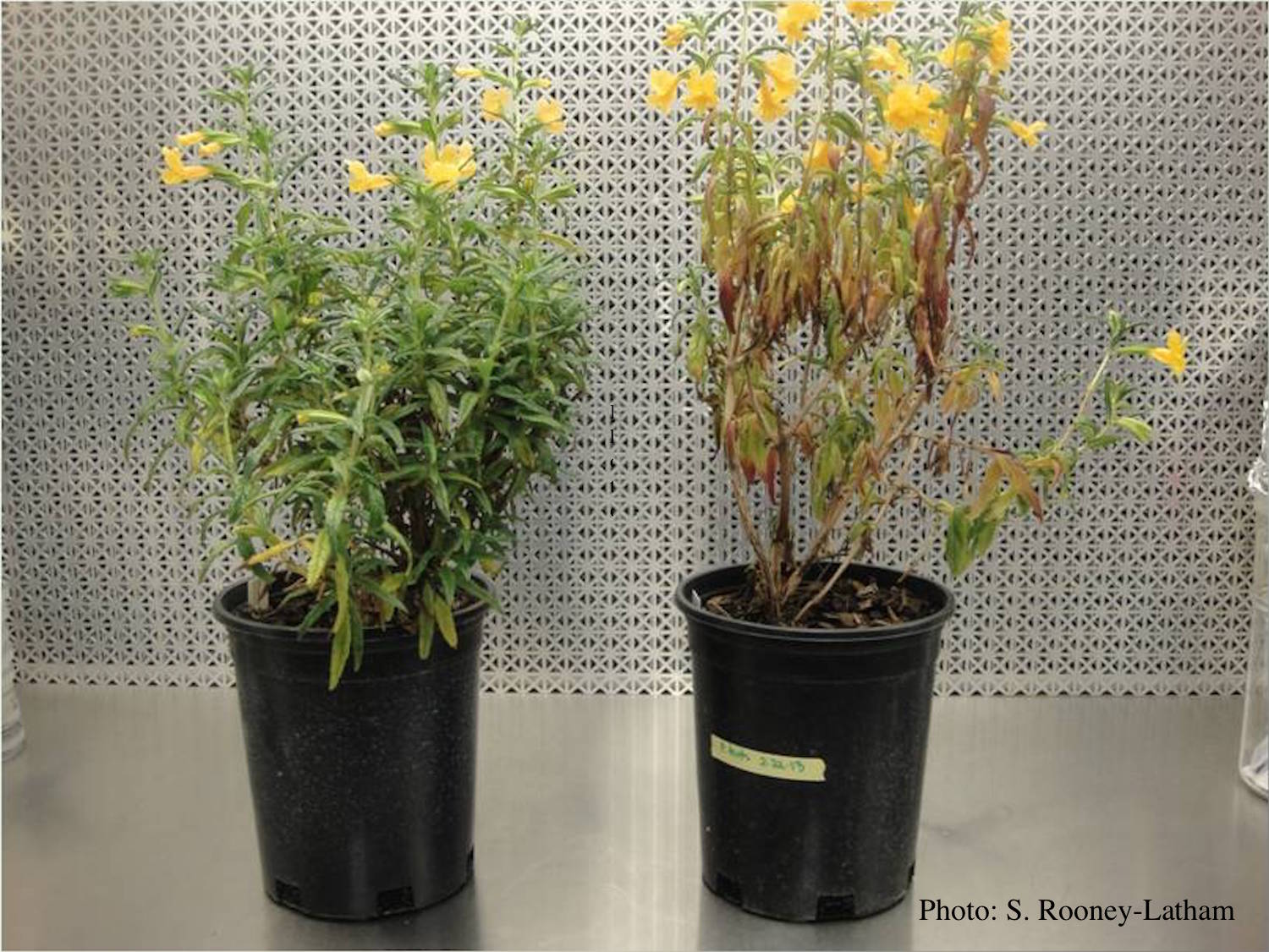 Crown and root rot (left) on sticky monkey flower (Diplacus aurantiacus) compared with a control (right) |
P. nicotianae sporangia  Noncaducous sporangium showing ovoid shape and papillate condition. (Fitopatol. bras. 2005) |
P. kernoviae sporangia  Mycol.Res 109, 853-859; Figs 18-22. Regular, ovoid limoniform sporangia. Figs 23-26. Asymmetrical or sporangia |