

Phytophthora nicotianae CBS 321.49 V8 after 7 days at 24 degrees. Photo from Q-bank: www.q-bank.eu, Henk Brouwer (CBS-KNAW, Utrecht, The Netherlands)
Photo Gallery
|
P. nicotianae colony morphology on V8 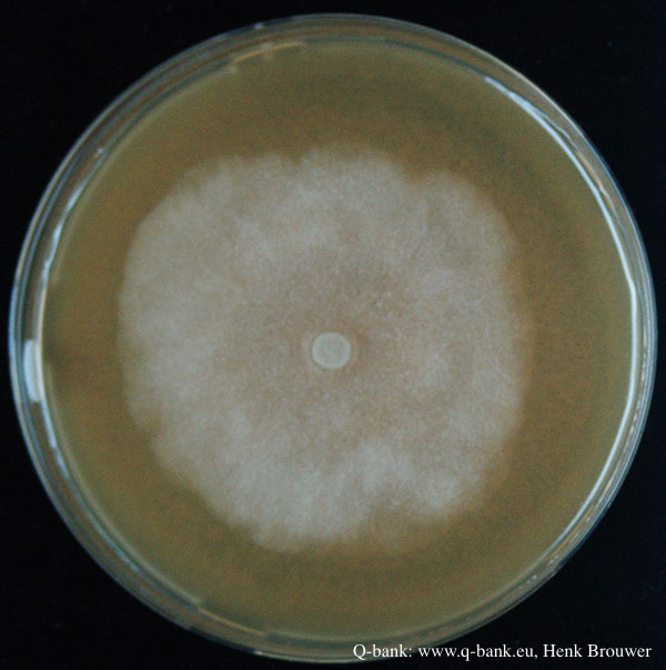 |
P. austrocedrae - hyphal swellings  Morphology of hyphae of Phytophthora austrocedrae, from Greslebin et al. 2007 |
P. cambivora disease symptoms 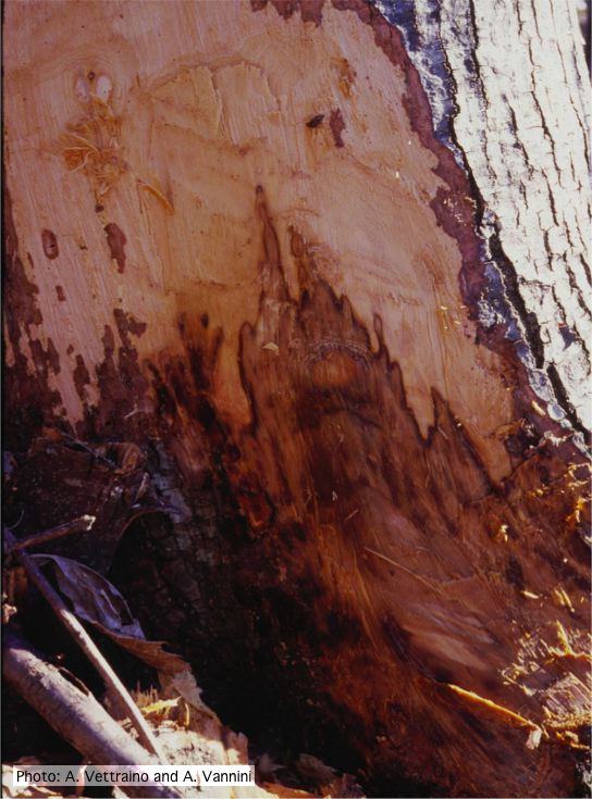 Collar canker rot of Ink disease on sweet chestnut |
|
P. cambivora bole canker 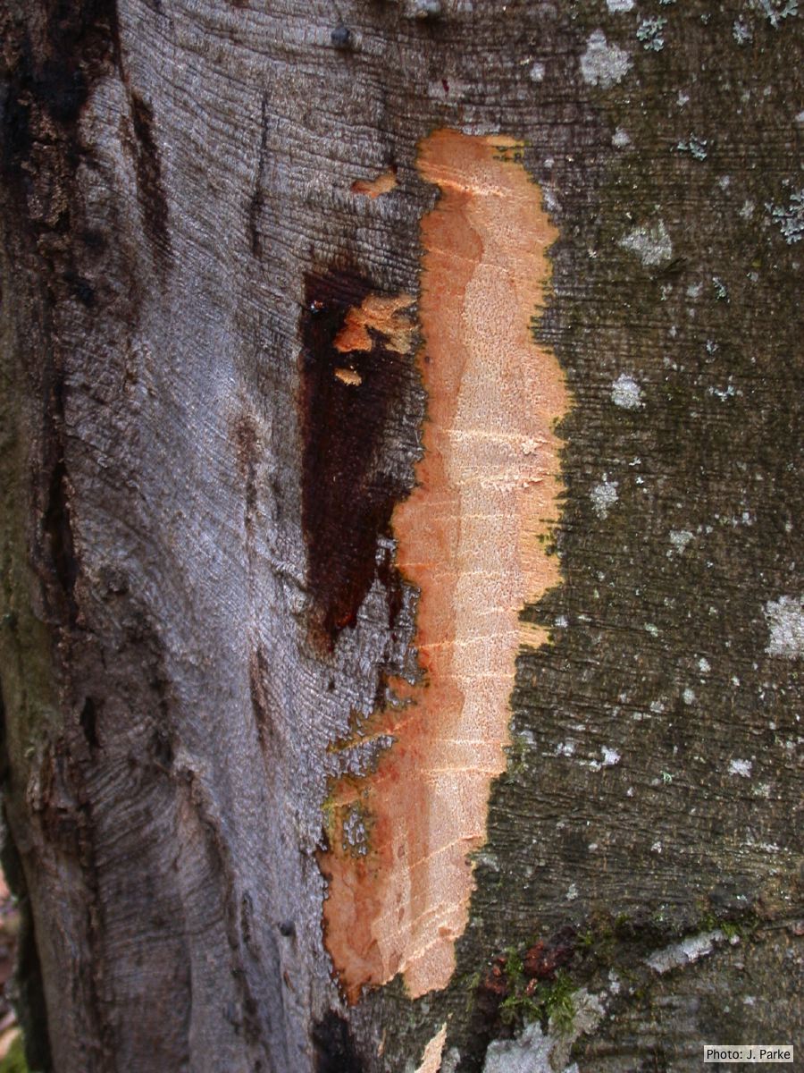 Fagus sylvatica bole canker |
P. chlamydospora chlamydospore 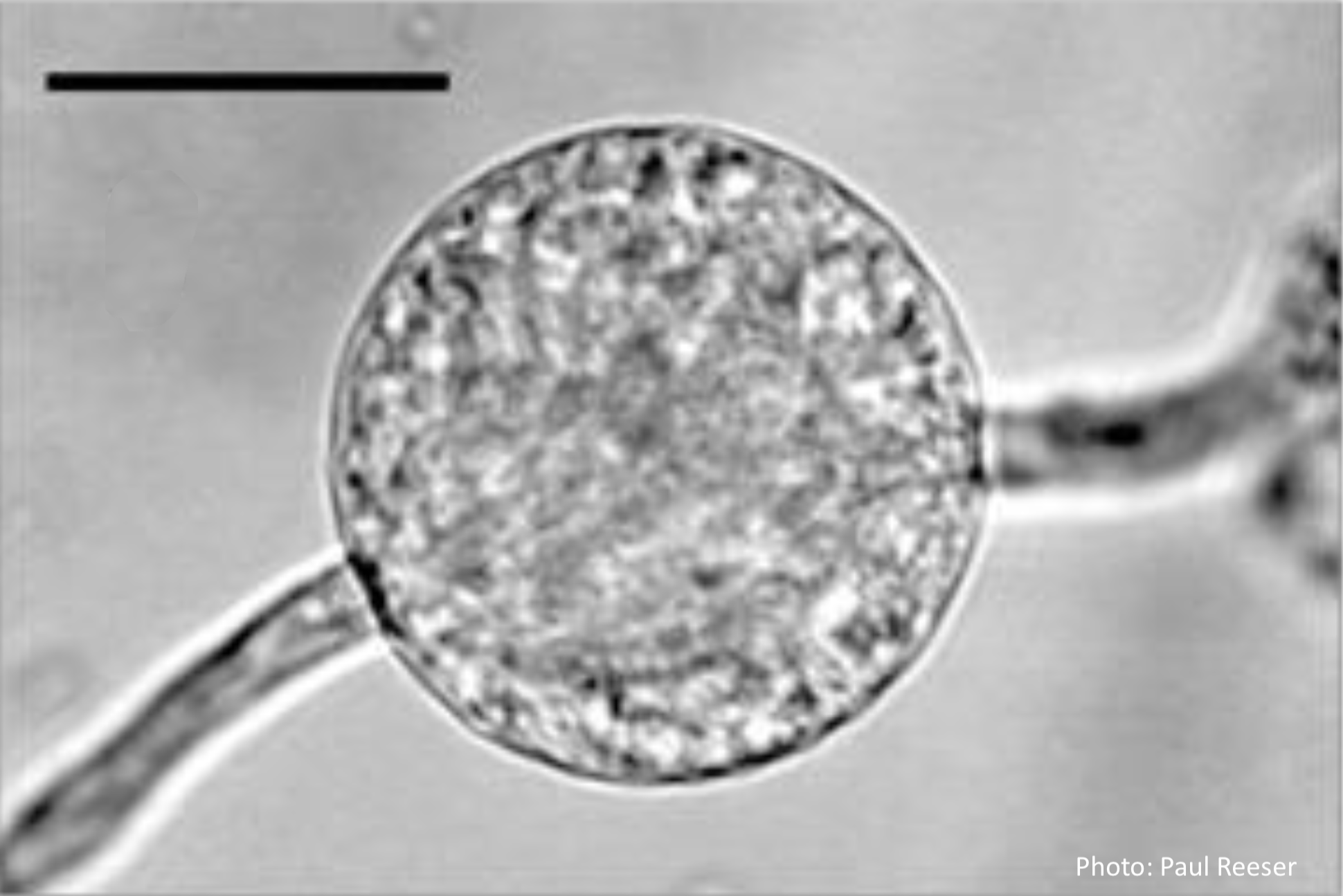 Phytophthora chlamydospora chlamydospore in agar. Bar is 20µm. |
P. pluvialis symptoms 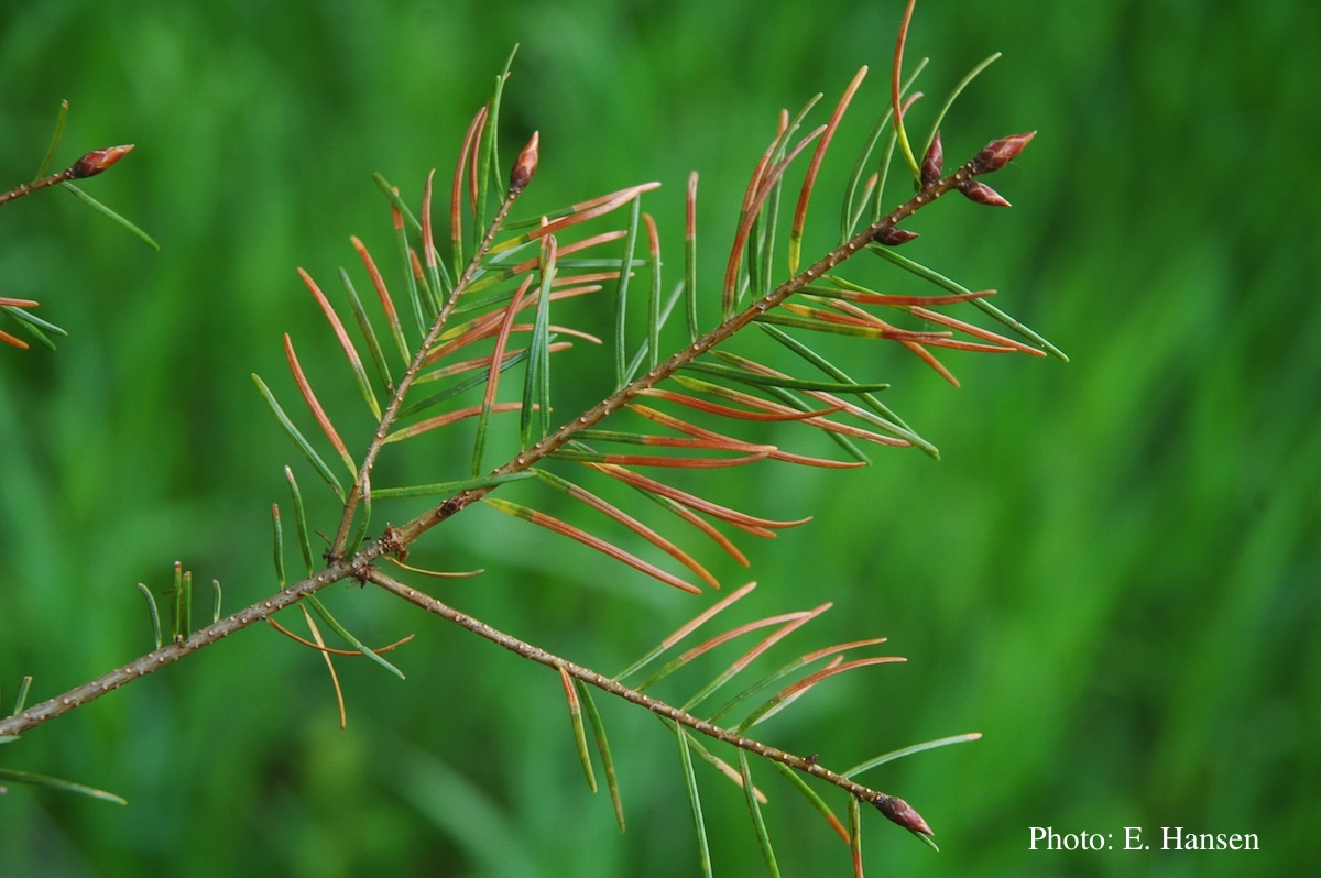 Symptoms of red needle cast on Douglas-fir needles |
|
P. palmivora symptoms on fruit  Brown rot on a lemon fruit caused by Phytophthora palmivora. |
P. chlamydospora colony morphology on carrot agar 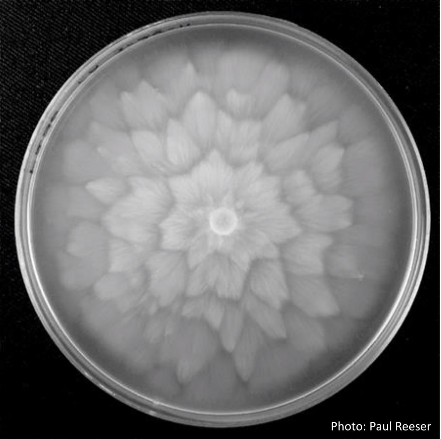 P. chlamydospora colony morphology on carrot agar |
P. katsurae oogonium 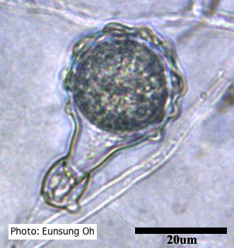 Warty protuberances on oogonium |
|
P. siskiyouensis bleeding canker  Bole lesions in the tissues under the bark of a bleeding canker: discoloration in the secondary phloem tissue |
P. kernoviae disease on European beech 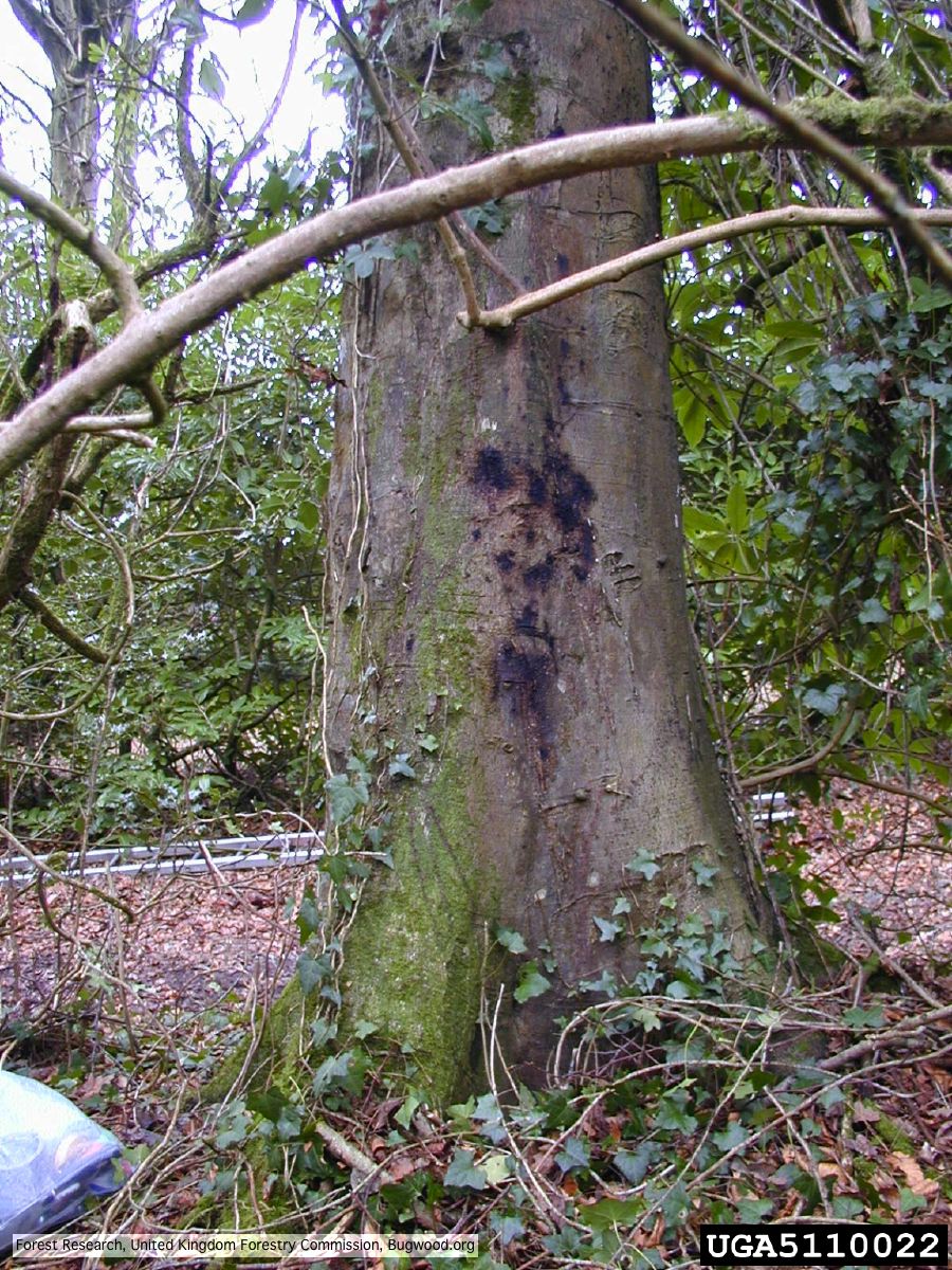 Bleeding lesion on trunk of Fagus sylvatica |
P. pluvialis symptoms on Douglas-fir 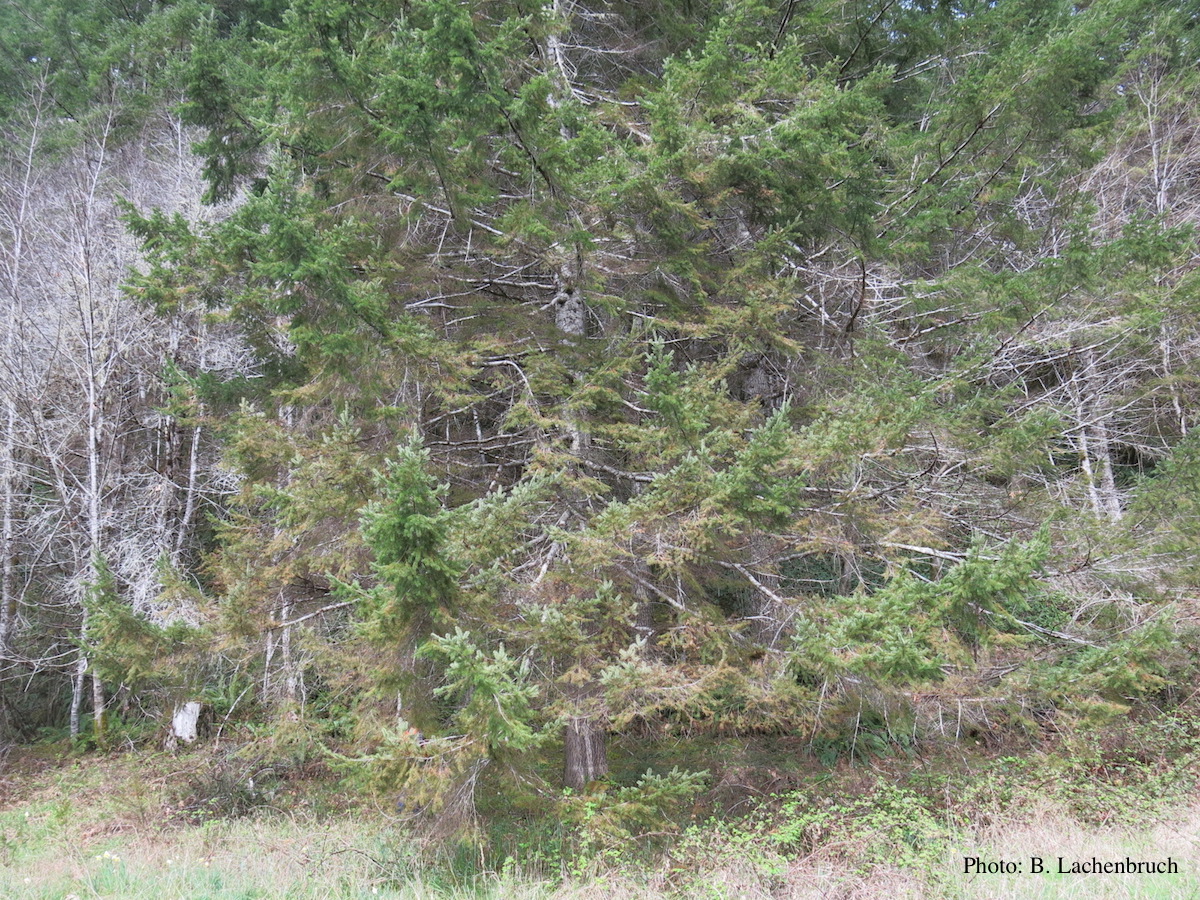 Red needle cast symptoms on Douglas-fir in western Oregon, 2015 |