

Papillate sporangium of P. tentaculata with an elongated neck or beak.
Photo Gallery
|
P. tentaculata sporangium 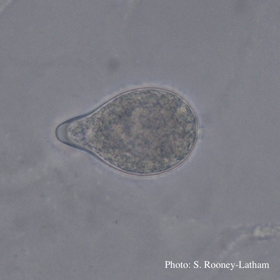 |
P. chlamydospora chlamydospore  Phytophthora chlamydospora chlamydospore in agar. Bar is 20µm. |
P. agathidicida lesion on kauri tree 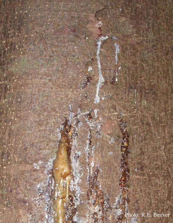 Gum oozing out of longitudinal lesion |
|
Basal canker on Port-Orford cedar 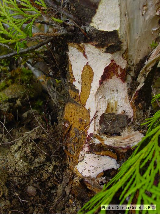 Basal canker on Chamaecyparis lawsoniana |
P. cambivora sporangium with internal extended proliferation  Empty sporagia showing internal nested and extended proliferation |
P. kernoviae sporangium 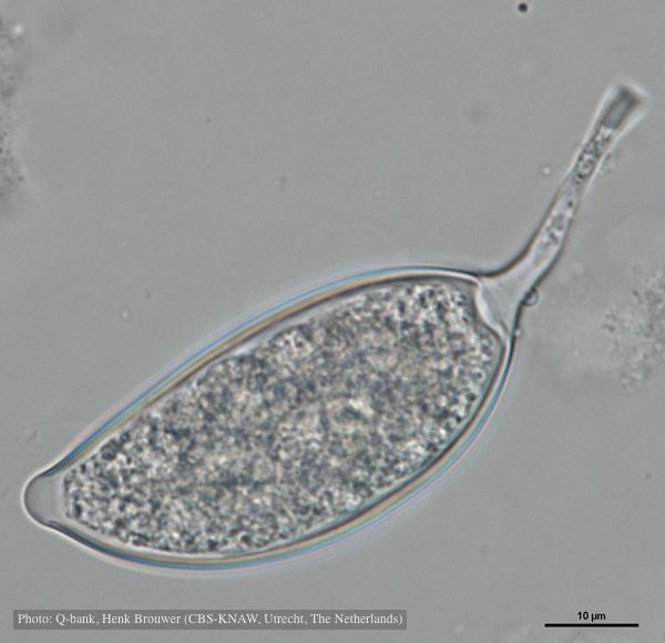 Asymmetrical sporangium, photo from Q-bank, used with permission |
|
P. tentaculata microscopic characteristics 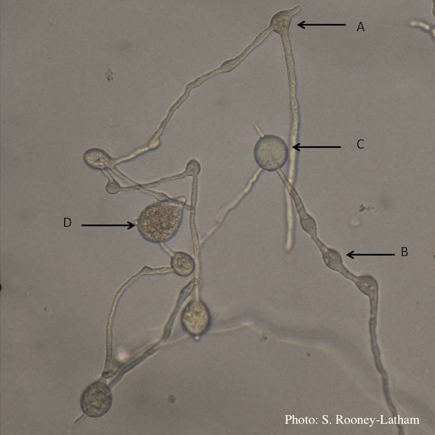 Hyphal swellings occuring at branching points of Mycelium (A), Intercalary hyphal swellings (B), Chlamydospore (C ), Sporangia (D) |
P. alni sporangia 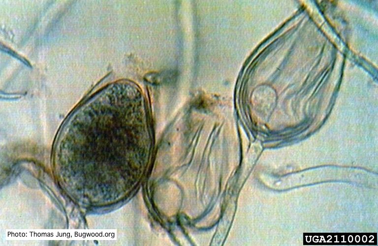 Non-papillate sporangia of P. alni showing nested proliferation. |
P. palmivora colony morphology on PDA 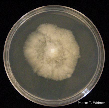 P. palmivora colony morphology on PDA |
|
Boot wash to station to control spread of P. agathidicida 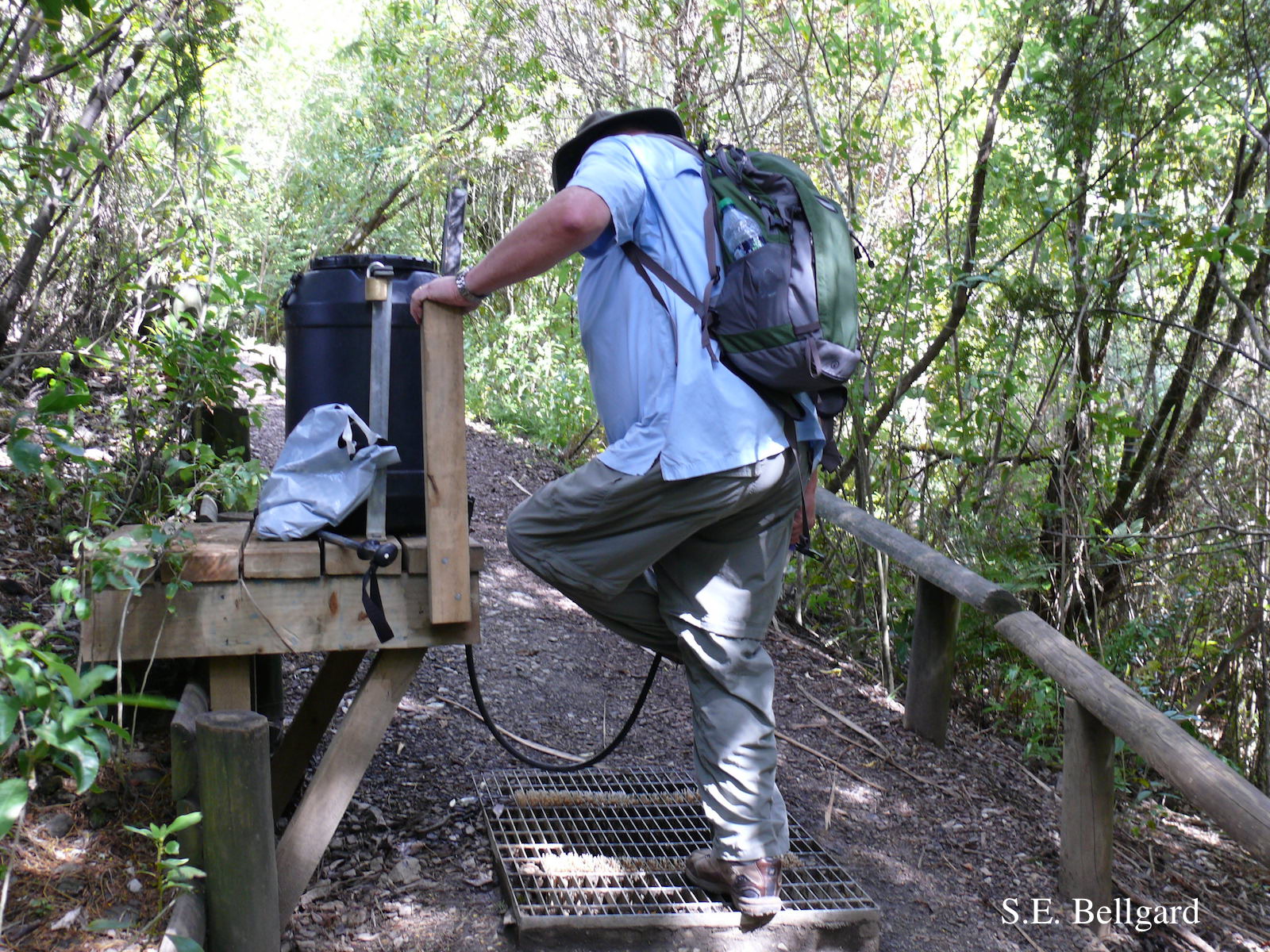 Use of hypochlorite solution applied through a “livestock drench-gun”, integrated with a soil grate to allow potentially contaminated soil to be collected |
P. pluvialis on Pinus radiata needle  Clusters of sporangia emerge from stomata of an infected radiata pine needle. |
P. kernoviae colony morphology  From Mycol.Res 109, 853-859; growth on CA under different conditions |