

Colony morphology on V8 at 7 days
Photo Gallery
|
P. megasperma colony morphology on V8  |
P. alni subsp alni sporangium 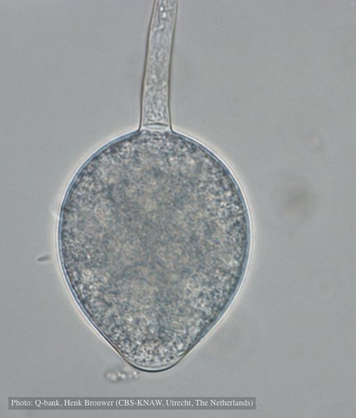 Non-papillate, non caducous sporangium, photo used with permission from Q-bank |
P. pinifolia sporangium 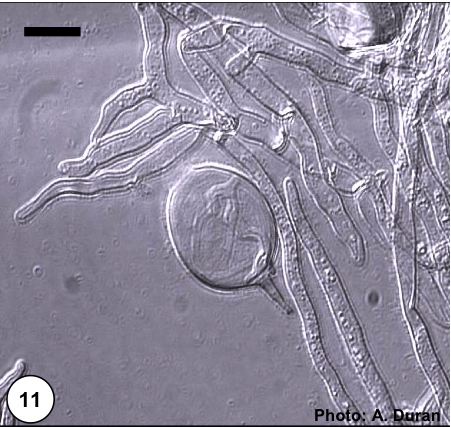 Non- papillate and caducous sporangia of Phytophthora pinifolia isolated from the infected P. radiata needles. |
|
Growth of P. arenaria on half-strength PDA 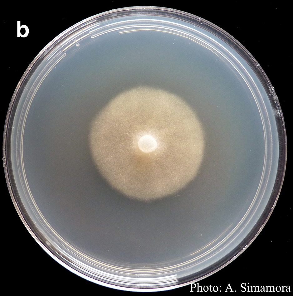 Colony morphology of Phytophthora arenaria after 7 days at 20°C on half-strength PDA |
P. ramorum oogonium 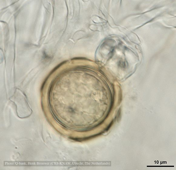 Oogonium with thick oogonial wall, photo from Q-bank, used with permission |
P. alni lesion in alder, Illwald, France 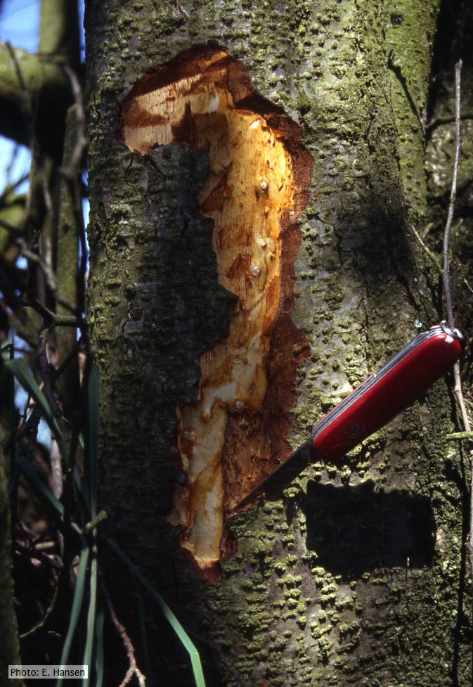 P. alni lesion in alder, Illwald, France |
|
P. pinifolia coenocytic hyphae 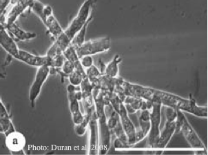 Coenocytic hyphae (from Duran et al. 2008). Scale bar = 20 μm. |
P. katsurae disease symptoms  Infected chestnut tree with girdling canker on stem |
P. ramorum bleeding canker 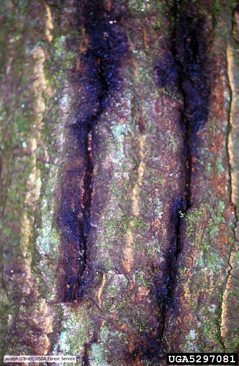 Bark cracks with black ooze in coast live oak, a symptom of P. ramorum canker. |
|
P. siskiyouensis canker on Italian alder 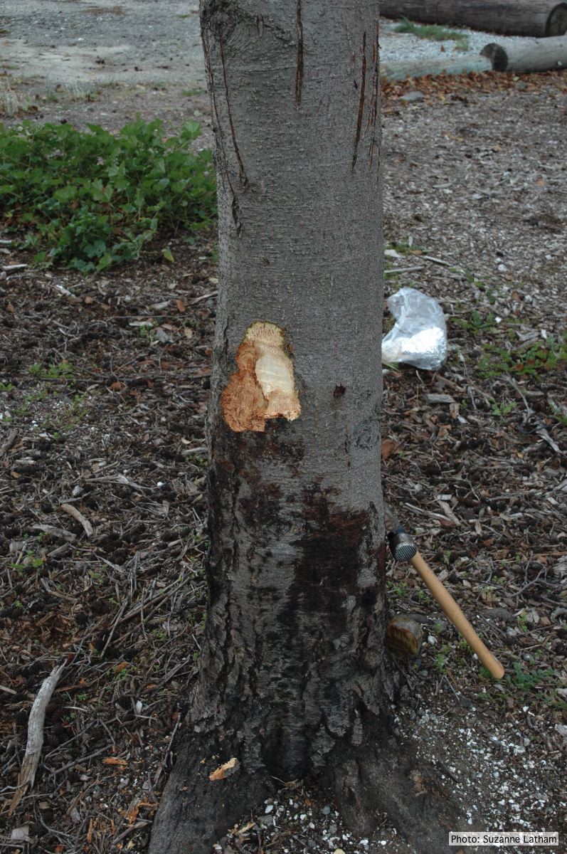 Bole lesions in the tissues under the bark of a bleeding canker: distinct margin between healthy and disease tissues |
P. cambivora disease symptoms 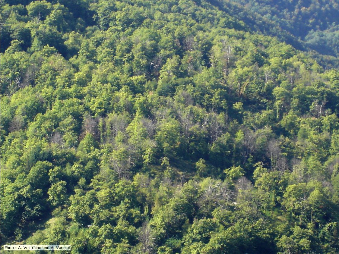 Ink disease impact in sweet chestnut forest in Italy |
P. cinnamomi cork oak decline  Cork oak decline, Portugal |