

Fig. 40. P. alni subsp. uniformis. Fig. 41. P. alni subsp. multiformis German variant. Fig. 42. P. alni subsp. alni. Fig. 43.
Photo Gallery
|
P. alni oogonia subspecies and variants 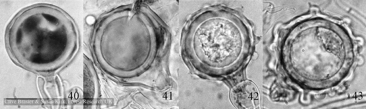 |
P. pseudosyringae oogonium  Oogonium with paragynous antheridium in agar |
P. lateralis sporangia 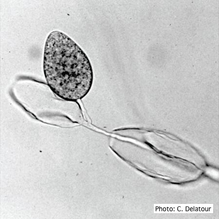 Sporangia of P. lateralis showing internal and external proliferation |
|
P. siskiyouensis bleeding canker  Bole lesions in the tissues under the bark of a bleeding canker: discoloration in the secondary phloem tissue |
P. kernoviae leaf wilt 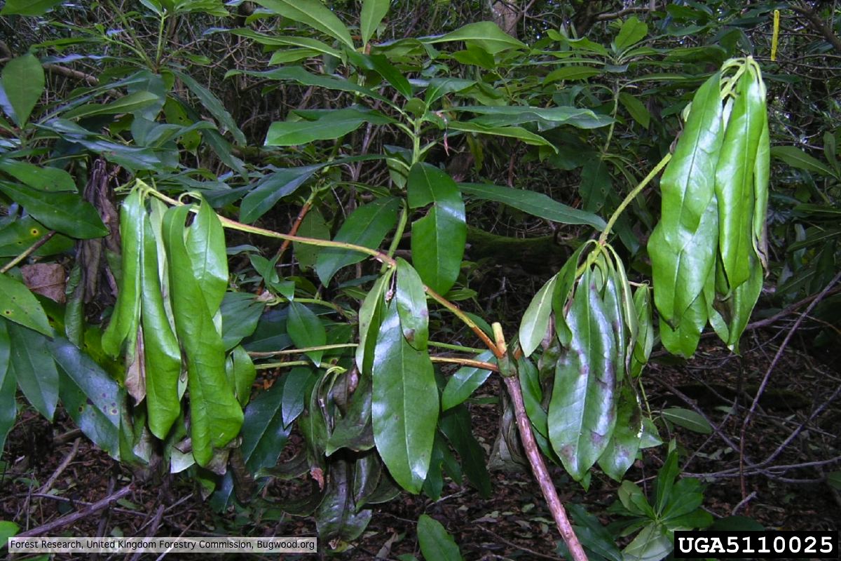 Wilted leaf of infected rhododendron |
P. megakarya disease symptoms on Theobroma cacao fruit 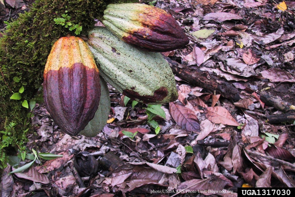 Disease symptoms on a cocoa pod |
|
P. austrocedrae colony morphology on Tomato juice agar 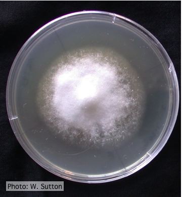 Colony morphology of P. austrocedrae at 16 ºC after 4 weeks on Tomato juice agar |
P. austrocedrae colony morphology on CMA 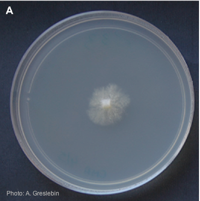 Colony morphology of P. austrocedrae at 16ºC after 4 weeks on CMA |
P. ramorum on larch in the United Kingdom 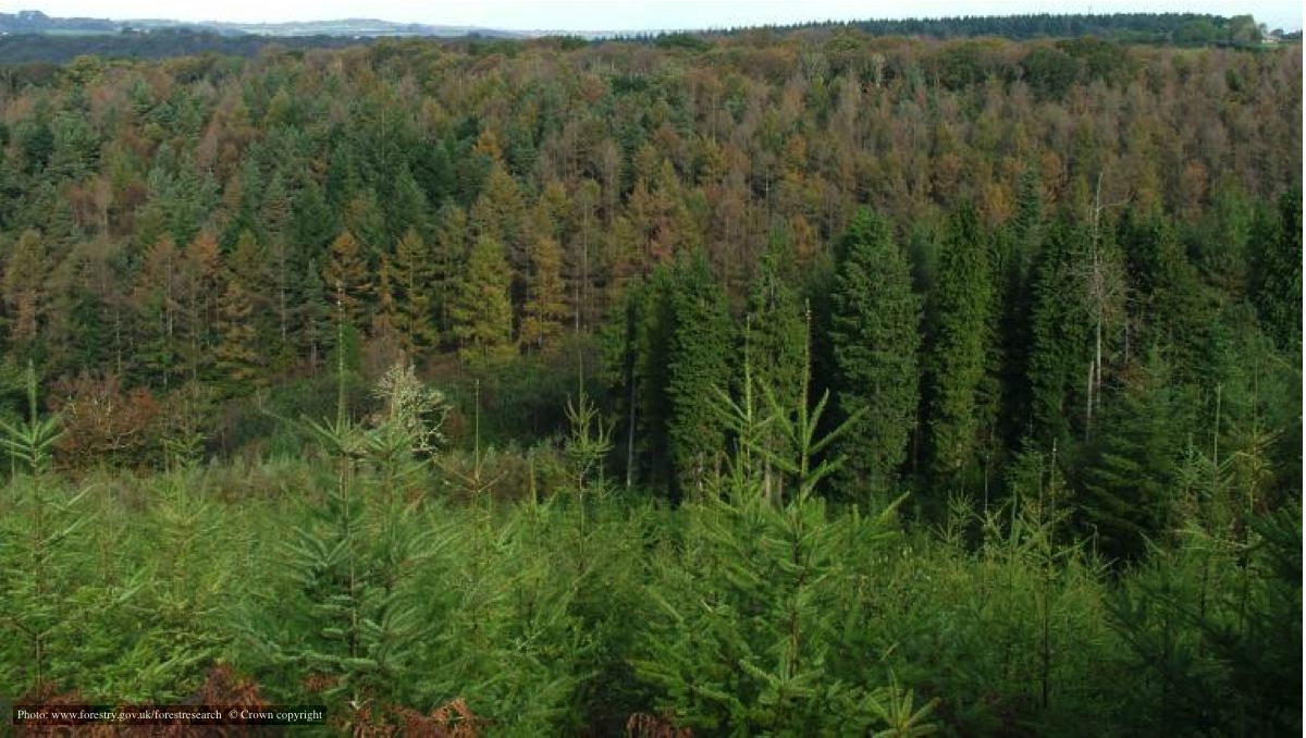 Larch trees with P. ramorum in the UK |
|
P. pluvialis symptoms on Douglas-fir 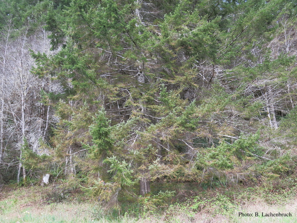 Red needle cast symptoms on Douglas-fir in western Oregon, 2015 |
P. pluvialis hyphal swellings 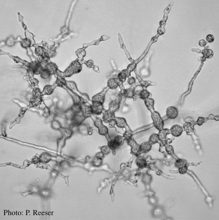 P. pluvialis hyphal swellings on agar |
P. cambivora colony morphology on MA 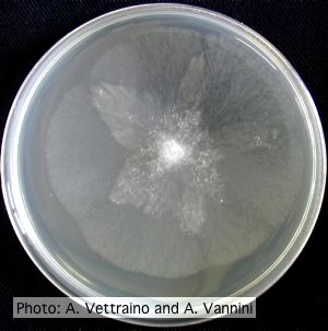 Appressed stellate colony morphology at 14 days at 20°C on MA |