

Pinus radiata needles, note “black line” symptom near needle bases
Photo Gallery
|
P. pinifolia on Pinus radiata 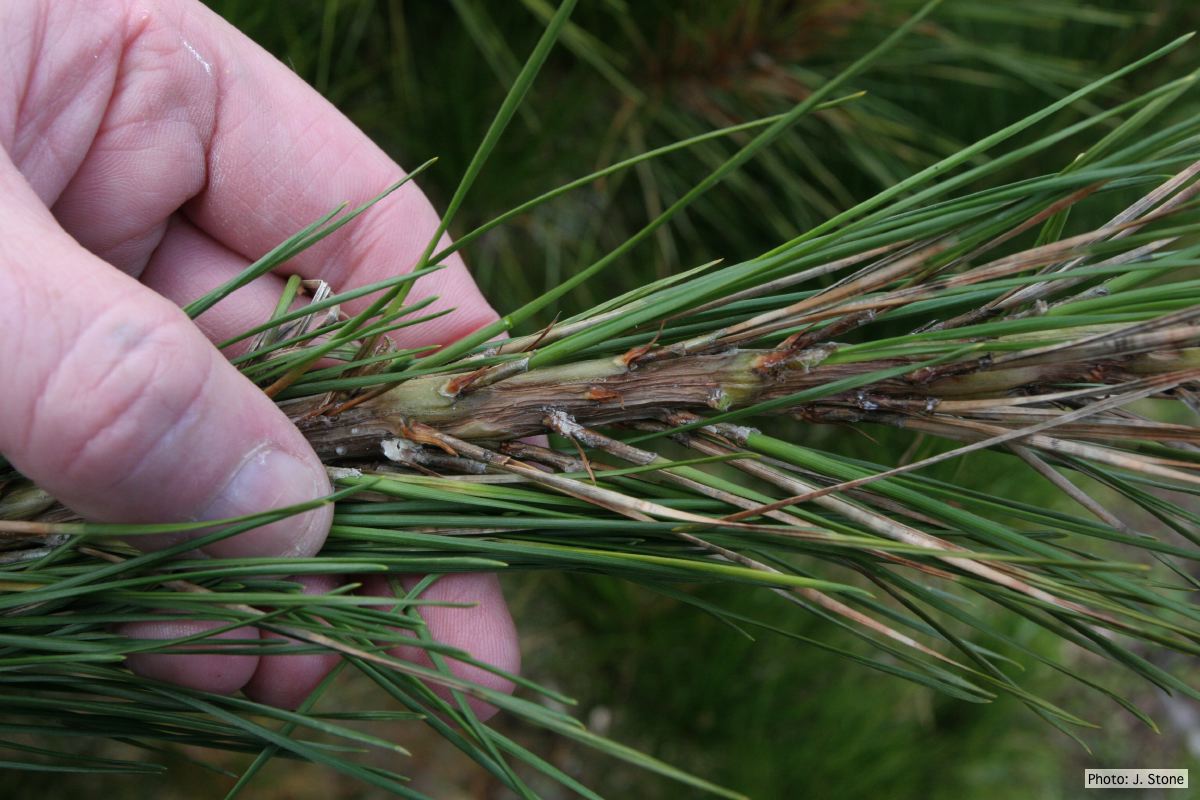 |
Mal del ciprés, dead and dying trees along river 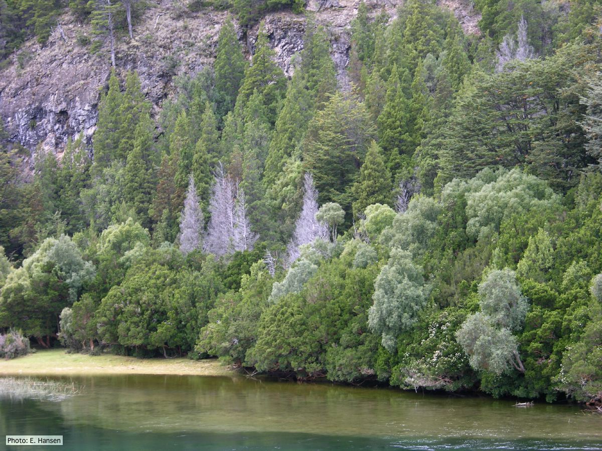 Mal del ciprés, dead and dying trees along river |
P. kernoviae disease on European beech 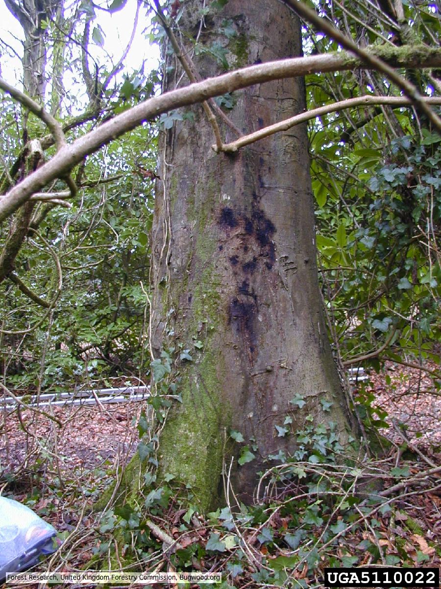 Bleeding lesion on trunk of Fagus sylvatica |
|
P. cactorum bleeding canker  Bleeding canker on European beech (Fagus sylvatica) |
P. siskiyouensis oogonium with amphigynous antheridium 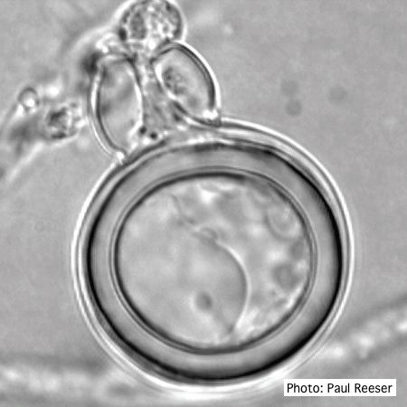 P. siskiyouensis oogonium with amphigynous antheridium |
P. chlamydospora colony morphology on V8 agar 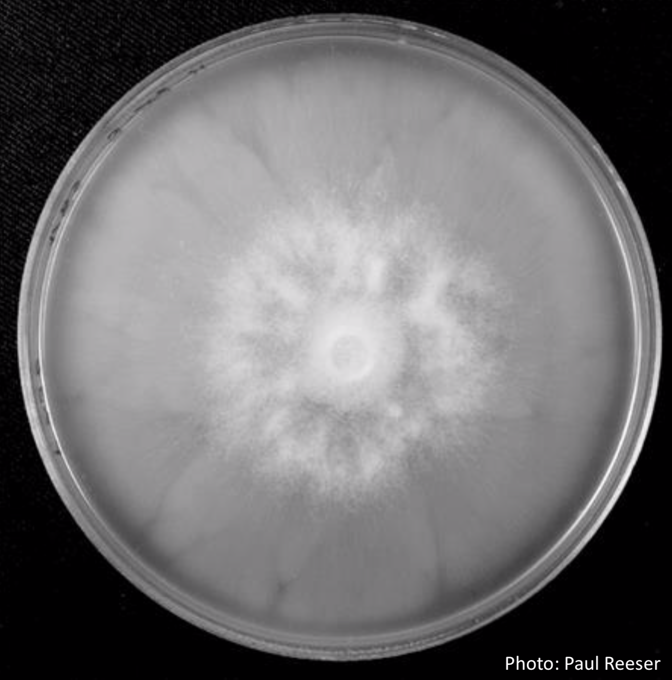 P. chlamydospora colony morphology on V8 agar |
|
Growth of P. arenaria on V8  Colony morphology of Phytophthora arenaria after 7 days at 20°C on V8 agar |
P. ramorum bleeding canker 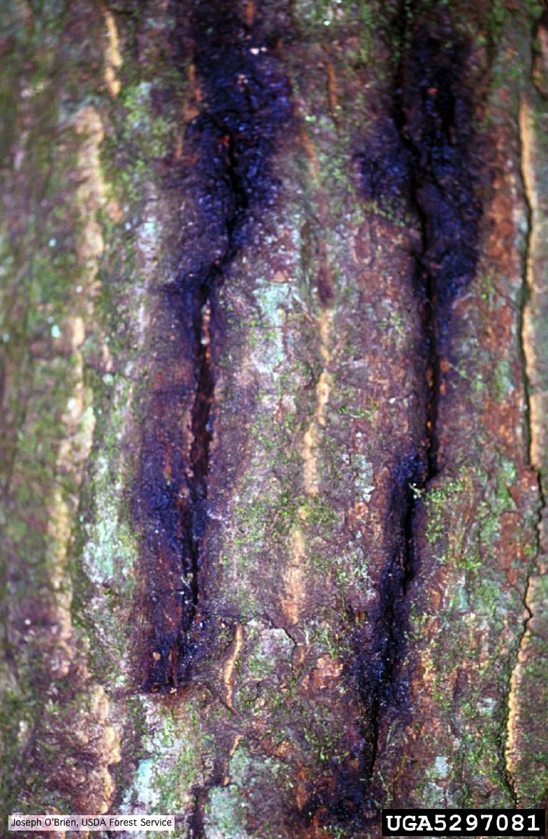 Bark cracks with black ooze in coast live oak, a symptom of P. ramorum canker. |
P. cambivora disease symptoms 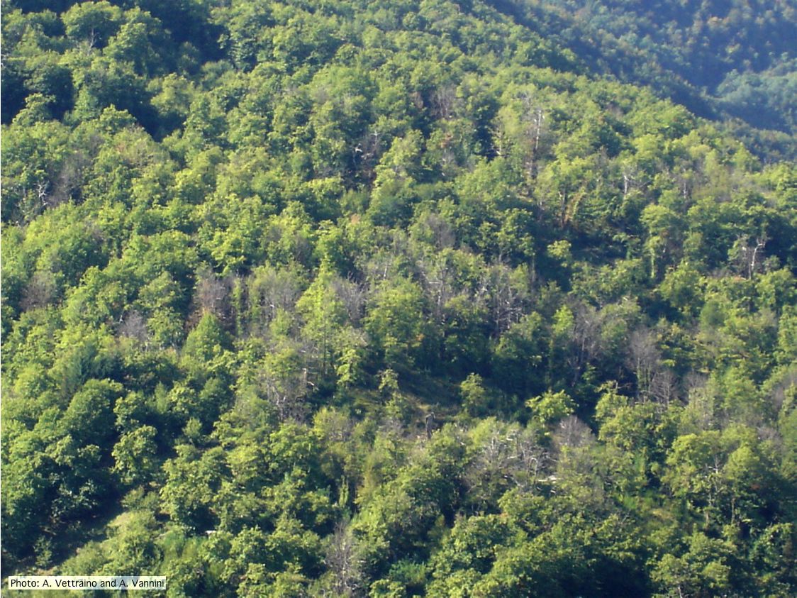 Ink disease impact in sweet chestnut forest in Italy |
|
P. cambivora oogonium 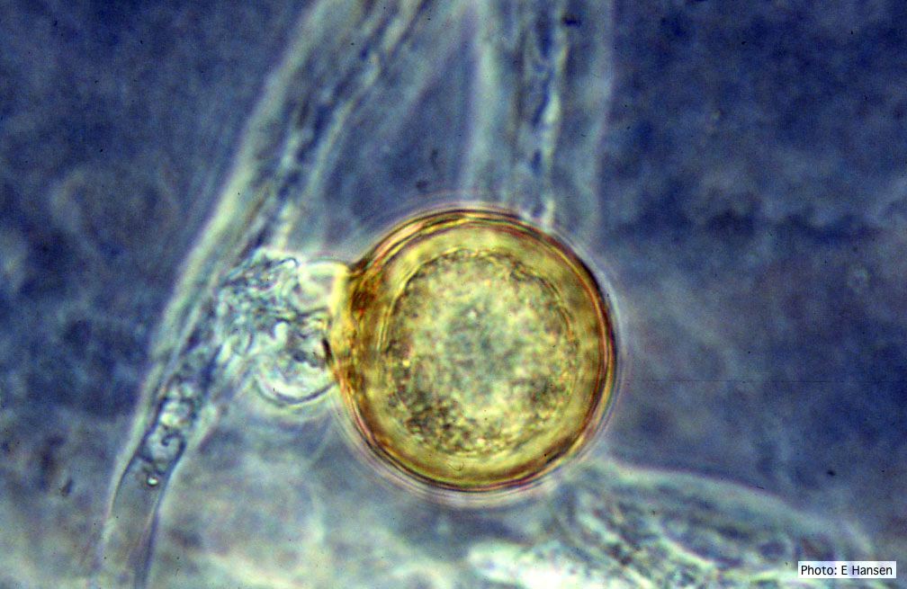 P. cambivora oogonium with antheridium |
P. cambivora colony morphology on PDA 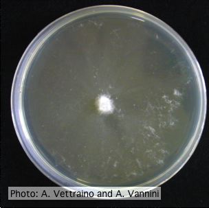 Appressed colony morphology at 14 days at 20°C on PDA |
Healthy Port Orford Cedar tree  Healthy Chamaecyparis lawsoniana trees |