

Sporangiophore showing internal proliferation through empty sporangia after zoospore release
Photo Gallery
|
P. cryptogea sporangia  |
P. pinifolia on Pinus radiata needles 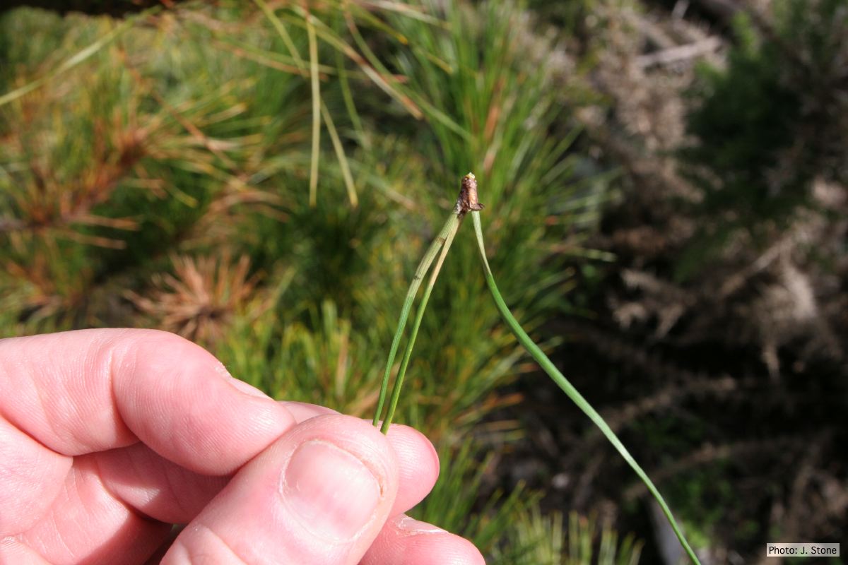 Pinus radiata needles, note “black line” symptom near needle bases |
P. pluvialis on Pinus radiata in New Zealand  Typical red needle cast symptoms along a twig. Lesions begin at the base of the needle which subsequently turns brown and is cast from the twig. |
|
P. ramorum sporangium  Deciduous sporangium, photo from Q-bank, used with permission |
P. austrocedrae oogonia drawing  P. austrocedrae. Morphology of oogonia, oospores and antheridia. Bar: 10 mm. Greslebin et al. 2007 |
P. megasperma oogonium 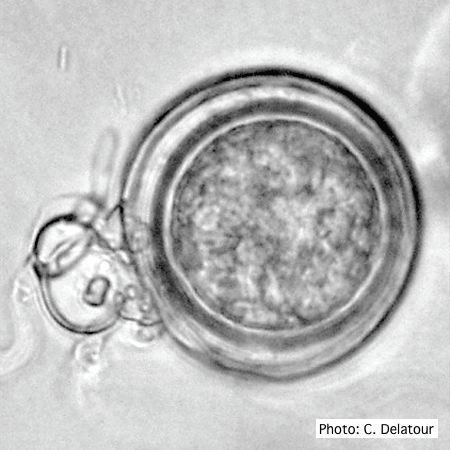 Oogonium with paragynous antheridia applied close to the ogonial stalk. |
|
P. nicotianae colony morphology on V8 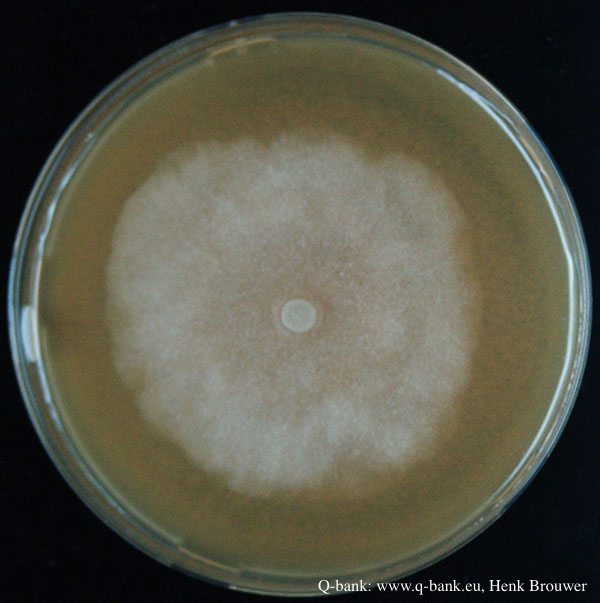 Phytophthora nicotianae CBS 321.49 V8 after 7 days at 24 degrees. Photo from Q-bank: www.q-bank.eu, Henk Brouwer (CBS-KNAW, Utrecht, The Netherlands) |
P. siskiyouensis oogonium with amphigynous antheridium 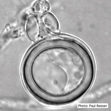 P. siskiyouensis oogonium with amphigynous antheridium |
P. pinifolia coenocytic hyphae 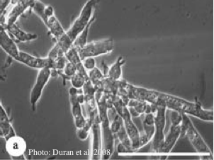 Coenocytic hyphae (from Duran et al. 2008). Scale bar = 20 μm. |
|
P. pinifolia on Pinus radiata  Pinus radiata, note Stem canker associated with necrotic needles. |
P. agathidicia sporangia 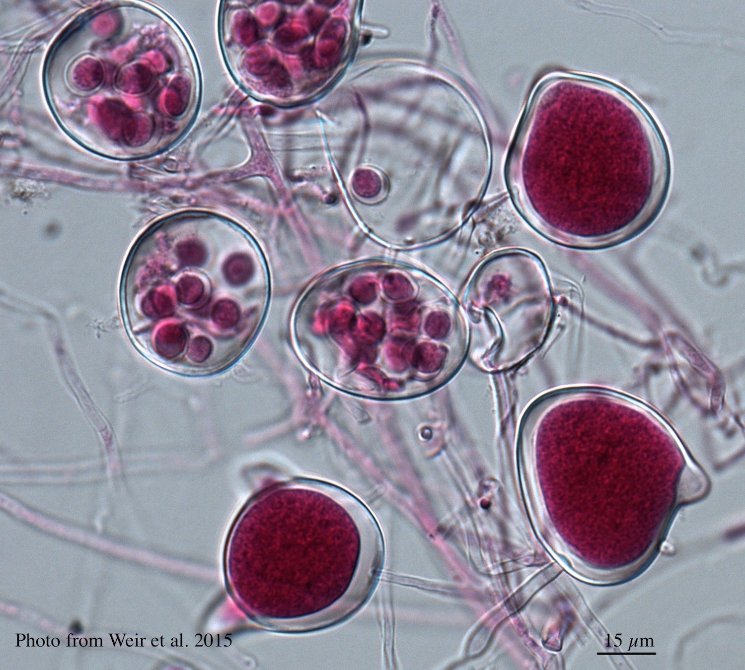 Differentiation of the cytoplasm within papillate sporangia into acid fuchsin stained zoospores |
P. lateralis on Port Orford cedar  Typical decline of Chaemacyparis lawsoniana in Landrévarzec, France |