

Bleeding canker on European beech (Fagus sylvatica)
Photo Gallery
|
P. cactorum bleeding canker 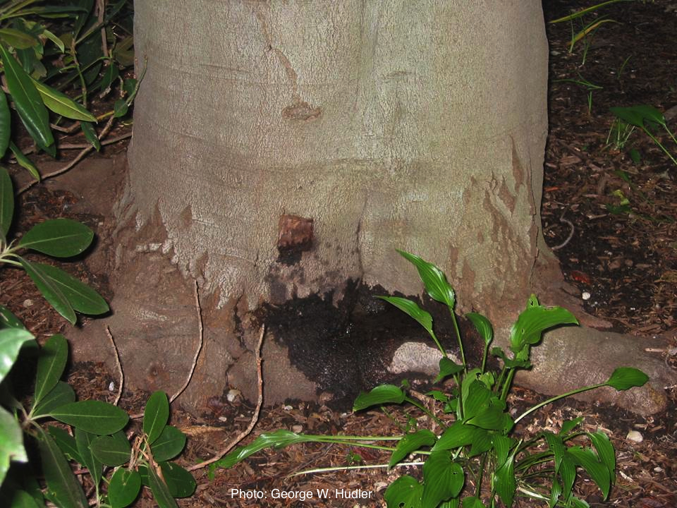 |
P. frigida symptoms 1 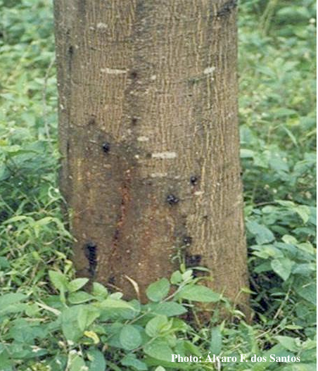 Symptoms of gummosis on black wattle |
P. megasperma sporangium  Ovoid, non-papillate sporangia |
|
P. pluvialis on Pinus radiata in New Zealand  Pinus radiata needles showing colour changes following infection with red needle cast disease. The tissues around the initial infection at the base or along the needle senesce, and change yellow and then brown as indicated by the arrows before the needles cast. |
P. cambivora on dead and dying chinquapin  Dead and dying chinquapin infected with P. cambivora |
Stain from Port Orford Cedar root disease  Stain from Chamaecyparis lawsoniana root disease on the Smith River |
|
P. chlamydospora hyphal swellings 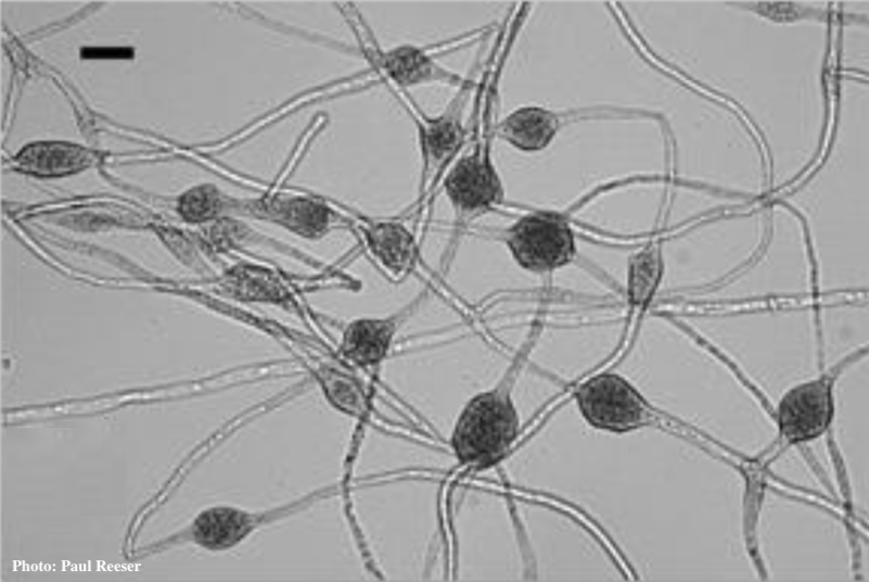 Phytophthora chlamydospora chlamydospore in agar. Bar is 20µm.
|
P. ramorum oogonium 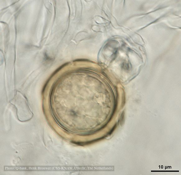 Oogonium with thick oogonial wall, photo from Q-bank, used with permission |
P. austrocedrae - hyphal swellings  Morphology of hyphae of Phytophthora austrocedrae, from Greslebin et al. 2007 |
|
P. pinifolia on Pinus radiata  Pinus radiata dead needles caused by DFP with healthy new growth from DFP |
P. cactorum bleeding canker  Bleeding canker on European beech (Fagus sylvatica) |
P. pluvialis symptoms 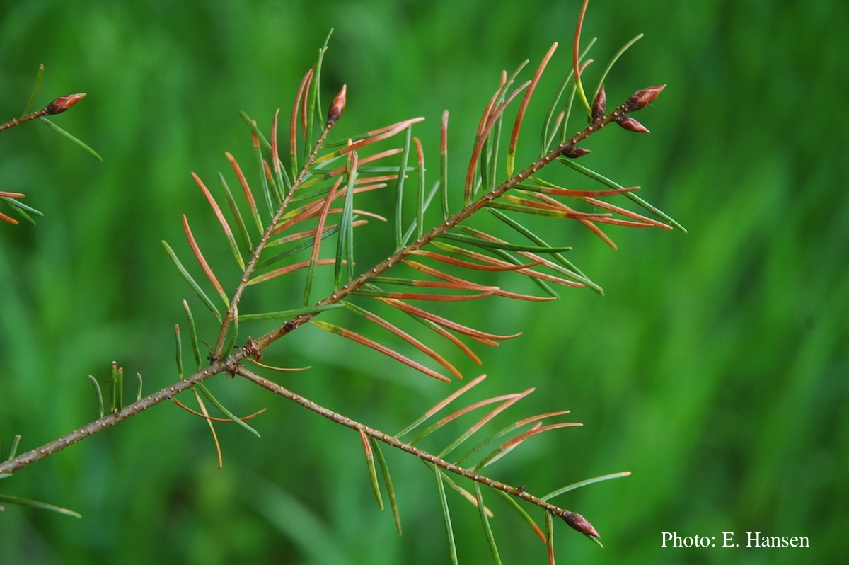 Symptoms of red needle cast on Douglas-fir needles |