

Canker on Fagus sp. caused by Phytophthora cactorum.
Photo Gallery
|
Phytophthora cactorum canker on beech tree  |
P. ramorum chlamydospores 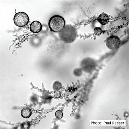 P. ramorum chlamydospores |
P. cambivora sporangium 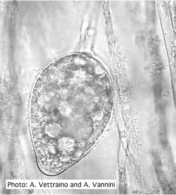 Ovoid non-papillate sporangia with well-rounded base |
|
P. nicotianae symptoms 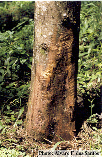 Symptoms of gummosis on black wattle (Fitopatol. bras. 2005) |
P. tentaculata microscopic characteristics 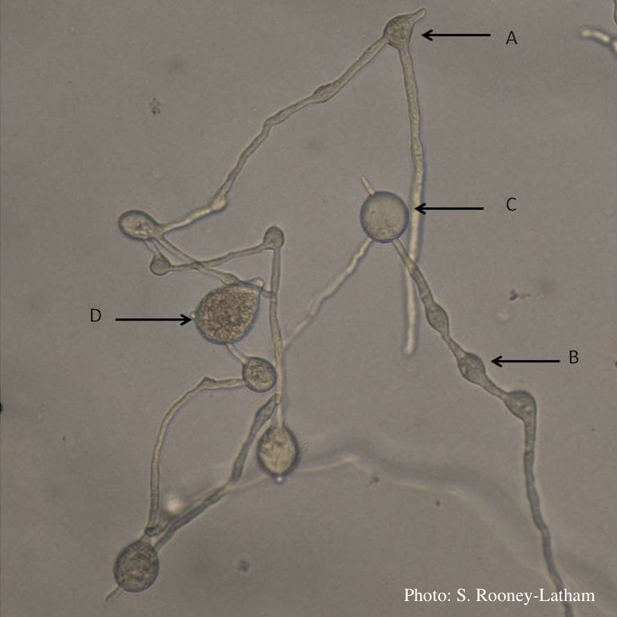 Hyphal swellings occuring at branching points of Mycelium (A), Intercalary hyphal swellings (B), Chlamydospore (C ), Sporangia (D) |
P. cambivora tar spots 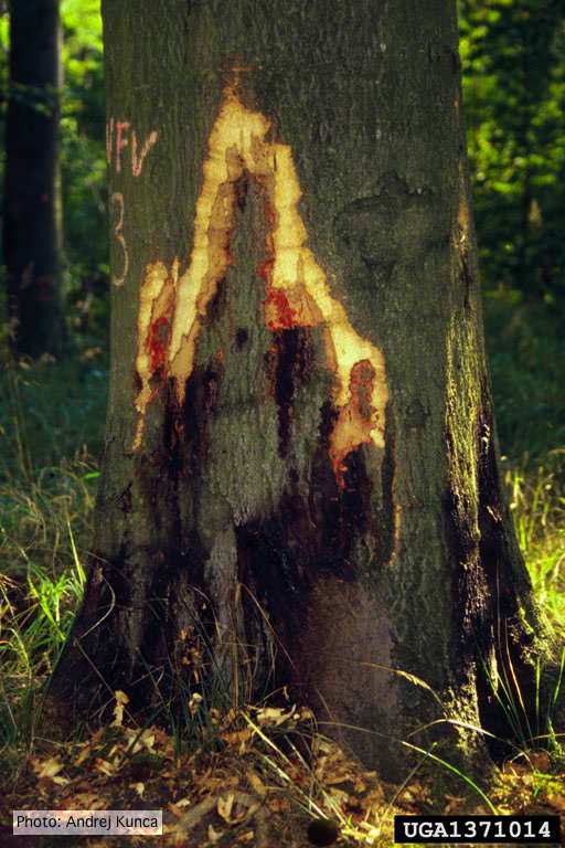 Tar spots on European beech (Fagus sylvatica) with bark removed. Lesse, Germany |
|
Growth of P. megakarya on corn meal agar
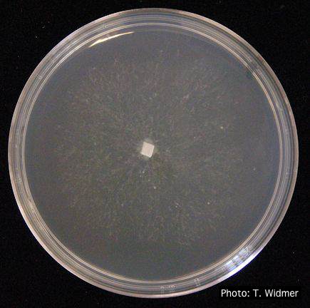 Growth of P. megakarya on corn meal agar |
P. megakarya disease symptoms on Theobroma cacao

Symptoms of black pod disease of cocoa (T. cacao)
|
P. pinifolia on Pinus radiata  Pinus radiata, note Infected needles at right angles to stem |
|
P. cambivora sporangium with nested proliferation 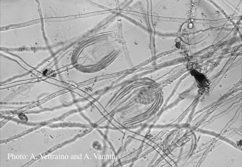 Empty sporagia showing internal nested proliferation |
P. tentaculata sporangium 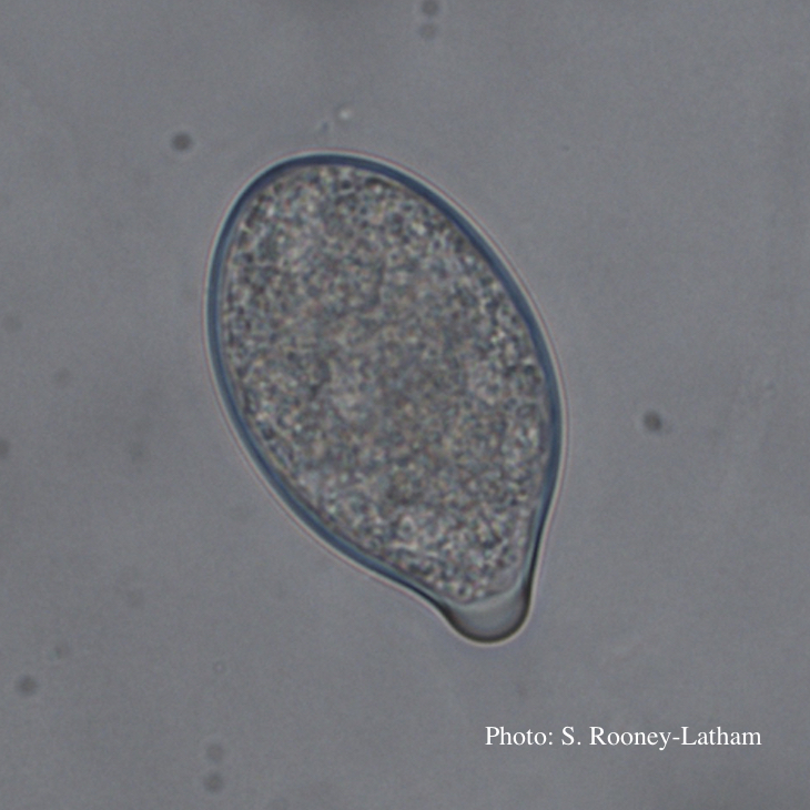 Papillate sporangium of P. tentaculata |
P. tentaculata disease symptoms on California mugwort 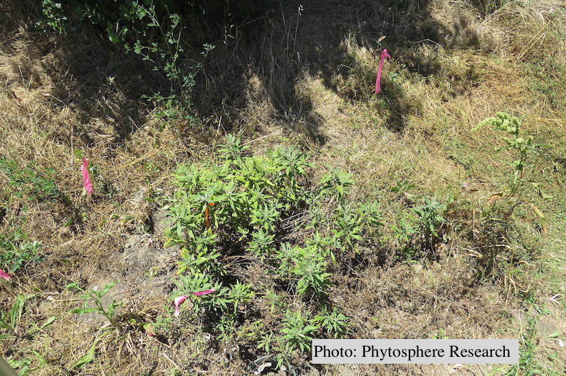 Outplanted California mugwort (Artemisia douglasiana) infected with P. tentaculata, 4.5 years after planting. Plant shows stunting and chlorosis. (P. cryptogea and P. lacustris were also baited from roots/soil of this plant). |