

Comparative gametangial morphology of Phytophthora Clade 5 species, with SEM (top) and light microscopy (bottom). P. heveae has smooth walled oogonia with funnel-shaped, amphigynous antheridia. P. agathidicida has mildly stipulate oogonia with globose amphigynous antheridia. P.cocois has mildly bullate oogonia with reflexed amphigynous antheridia. P. castaneae has coarsely bullate oogonium with rugose protuberances and narrow amphigynous antheridia (Weir et al. 2015).
Photo Gallery
|
Comparative gametangial morphology of Phytophthora Clade 5 species  |
P. cambivora sporangium 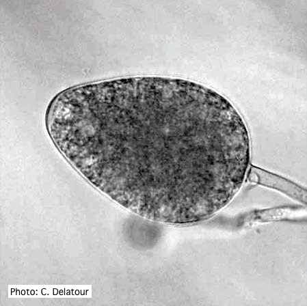 Ovoid non- papillate sporangia |
P. nicotianae colony morphology on PDA 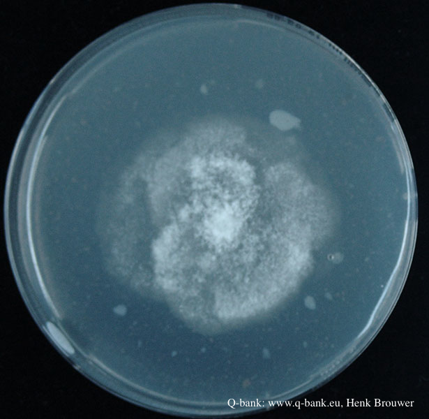 Phytophthora nicotianae CBS 321.49 PDA after 7 days at 24 degrees. Photo from Q-bank: www.q-bank.eu, Henk Brouwer (CBS-KNAW, Utrecht, The Netherlands) |
|
P. kernoviae sporangium 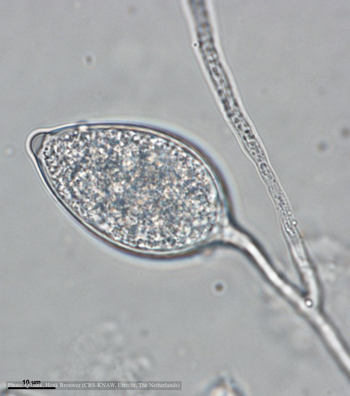 Papillate and caducous sporangium, photo from Q-bank, used with permission |
P. ramorum leaf symptoms on tan oak 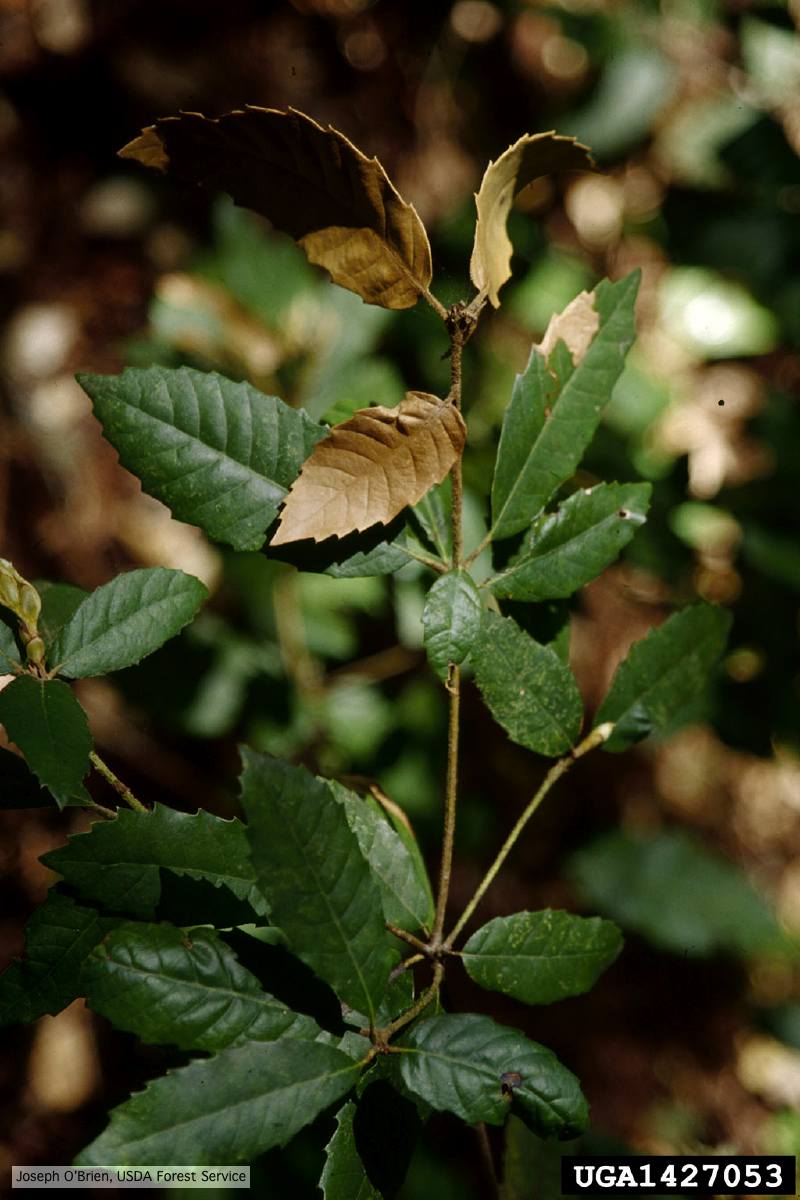 Tip symptoms on tanoak seedling (Notholithocarpus densiflorus). |
P. austrocedrae hyphal swellings 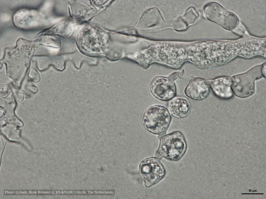 Hyphal swelling photo used with permission from Q-bank |
|
P. arenaria disease symptoms on Banksia landscape 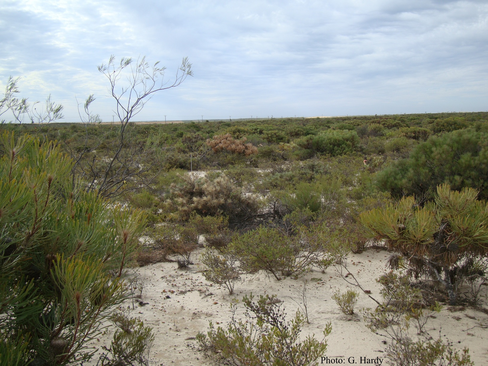 Dead Banksia sp. in a Kwongan heathland on mineral sand near Eneabba, Western Australia recently killed by root and collar rot caused by Phytophthora arenaria |
P. agathidicia sporangia 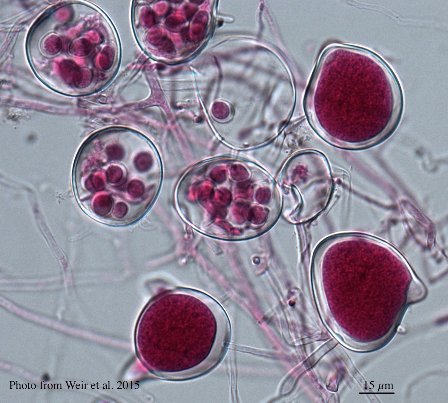 Differentiation of the cytoplasm within papillate sporangia into acid fuchsin stained zoospores |
P. megasperma sporangium  Ovoid, non-papillate sporangia |
|
P. palmivora sporangia 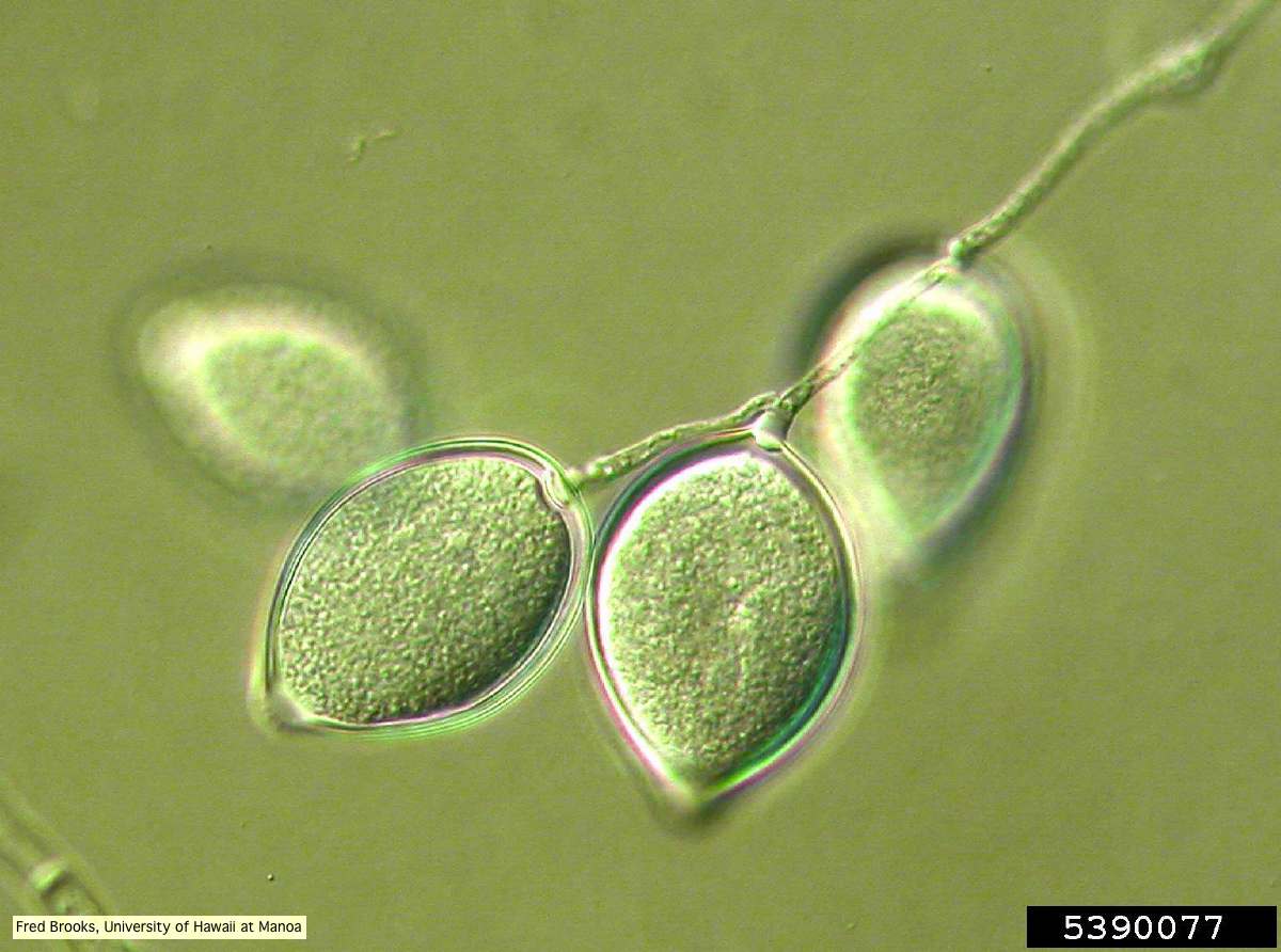 Sporangia (sporangiospores) showing sympodial branching |
P. pluvialis symptoms on Douglas-fir 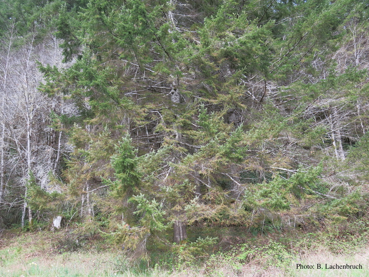 Red needle cast symptoms on Douglas-fir in western Oregon, 2015 |
P. cambivora sporangia 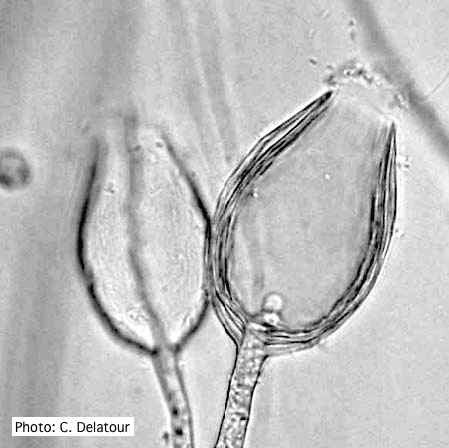 Empty sporangia of P. cambivora showing nested internal proliferation |