

Ovoid, semipapillate sporangia showing medium length pedicel
Photo Gallery
|
P. pseudosyringae sporangium 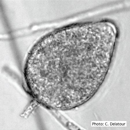 |
P. katsurae oogonia 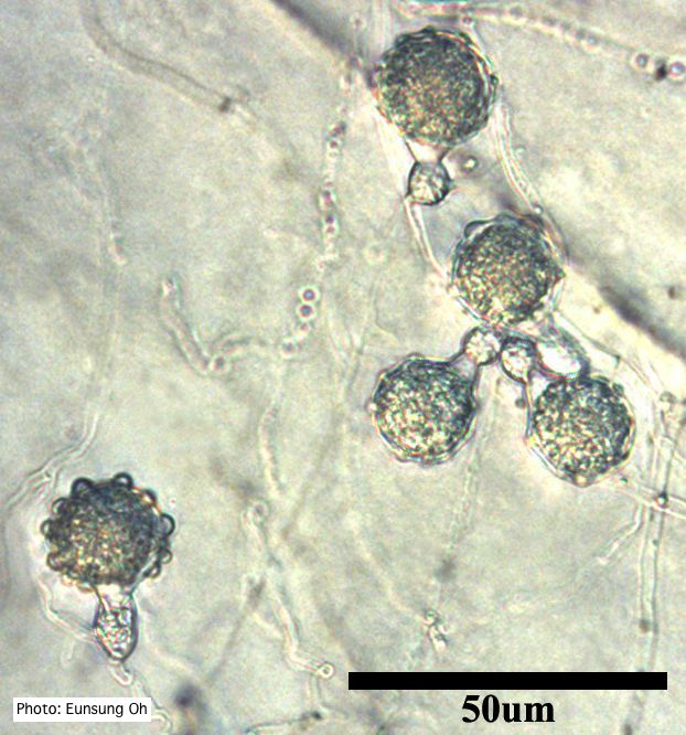 Oogonia with ornamentation |
P. cambivora oogonium 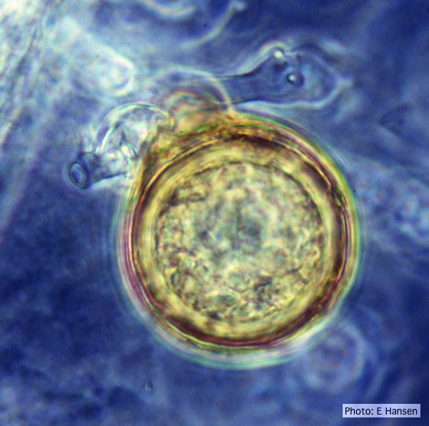 P. cambivora oogonium with antheridium |
|
P. frigida symptoms 4 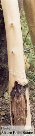 Black wattle timber with symptoms of gummosis |
P. austrocedrae - Mal del ciprés, stages of decline 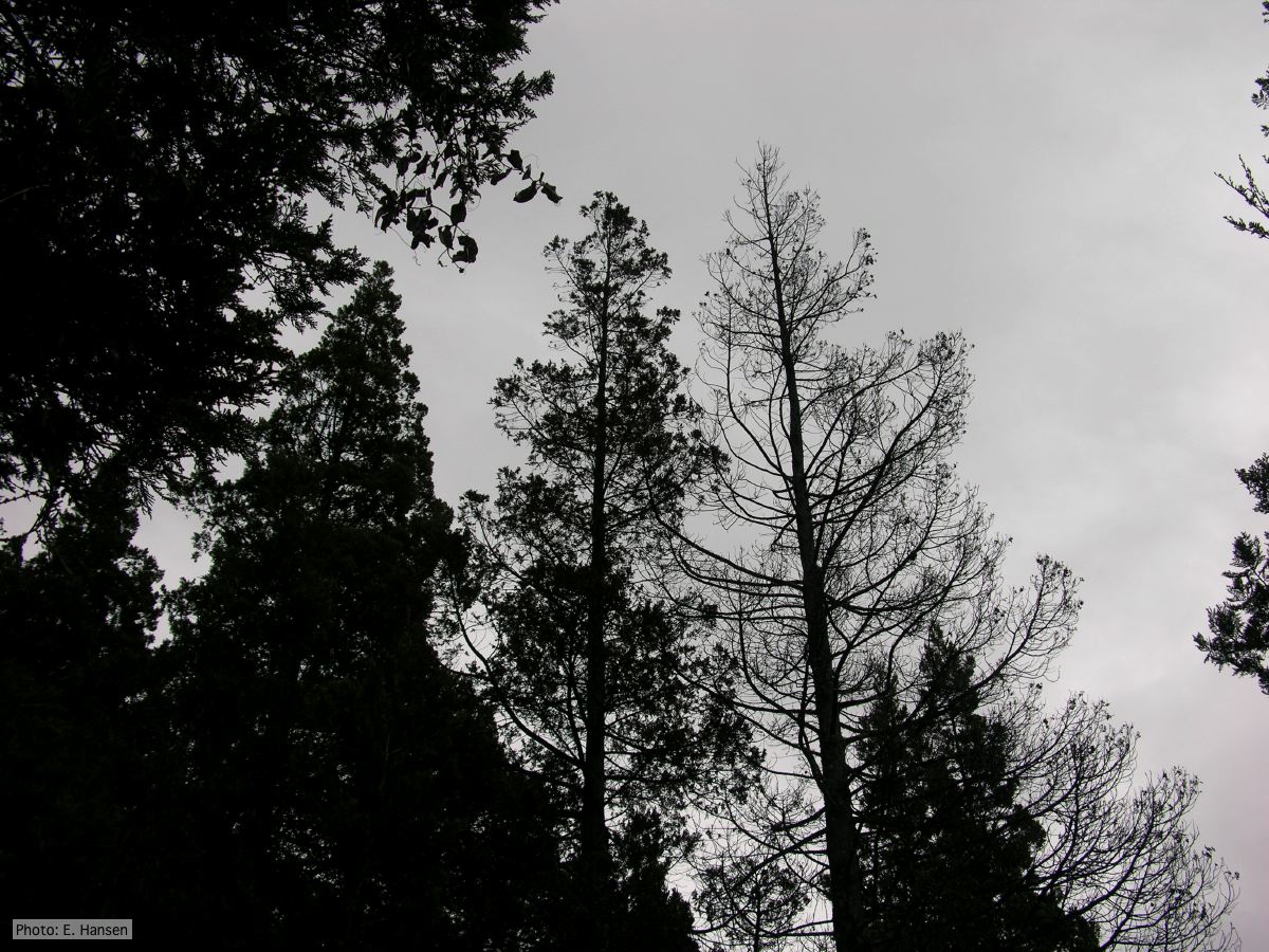 Mal del ciprés, stages of decline |
Port Orford Cedar Hedge row  Chamaecyparis lawsoniana residential hedge row with alive and dead trees |
|
P. tentaculata disease symptoms on sticky monkey flower 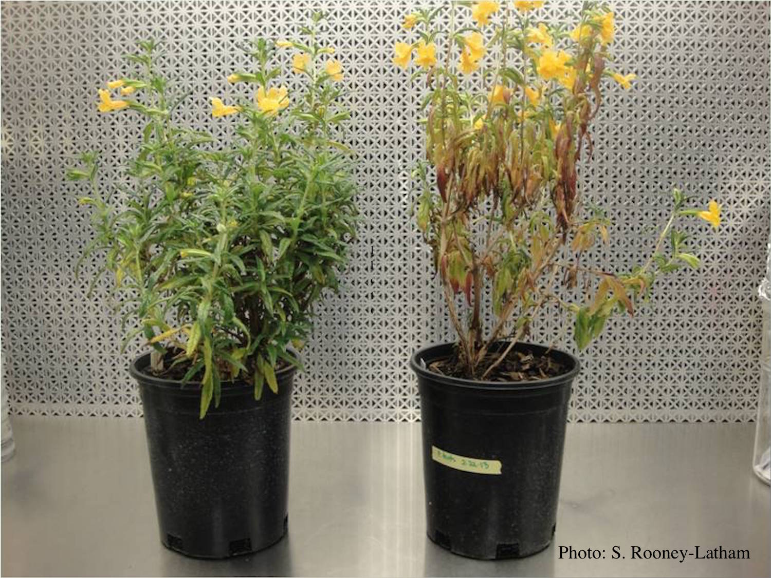 Crown and root rot (left) on sticky monkey flower (Diplacus aurantiacus) compared with a control (right) |
P. pluvialis on Pinus radiata 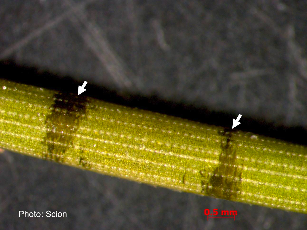 A Pinus radiata needle showing black resinous bands or marks consistent with the presence of red needle cast disease. |
P. pluvialis on Pinus radiata in New Zealand 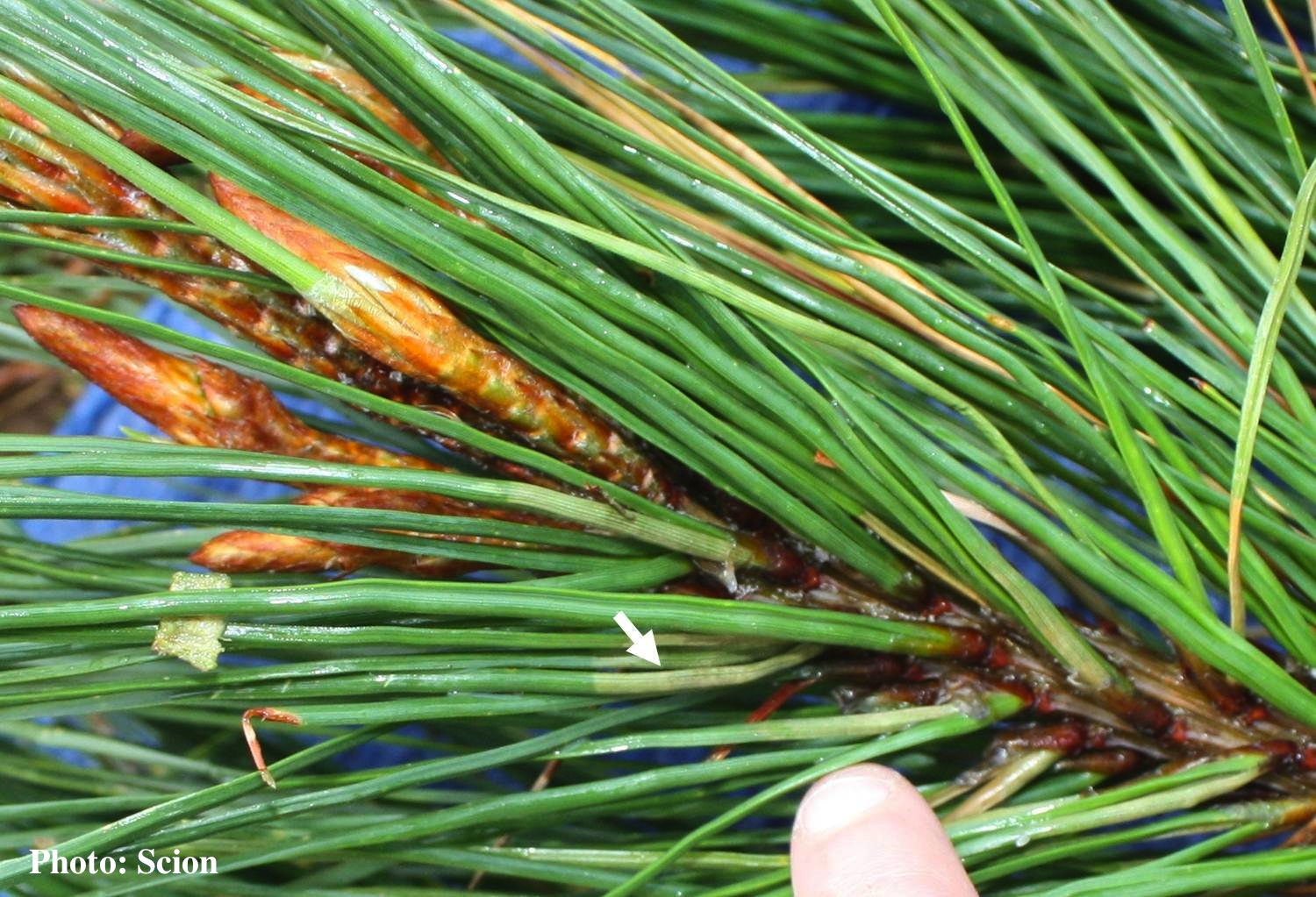 Lesions consistent with the presence of red needle cast disease are more abundant at the base of Pinus radiata needles as indicated by the arrow. |
|
P. palmivora chlamydospore 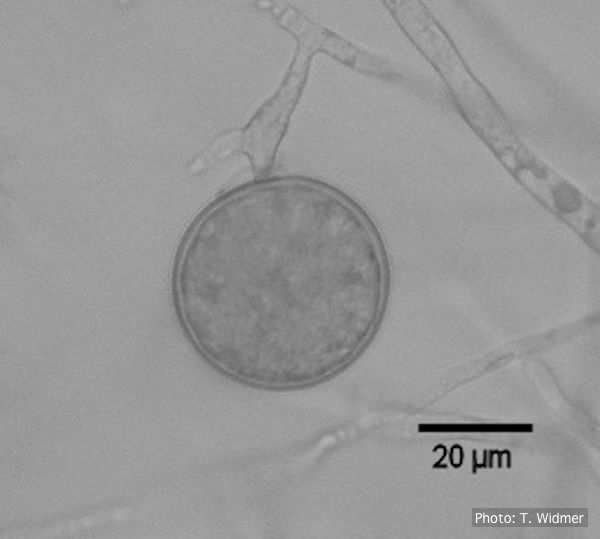 Terminal chlamydospore of P. palmivora |
P. arenaria oogonium  Aplerotic oogonia of P. arenaria with paragynous antheridia. Scale bar = 20 μm |
P. chlamydospora sporangium 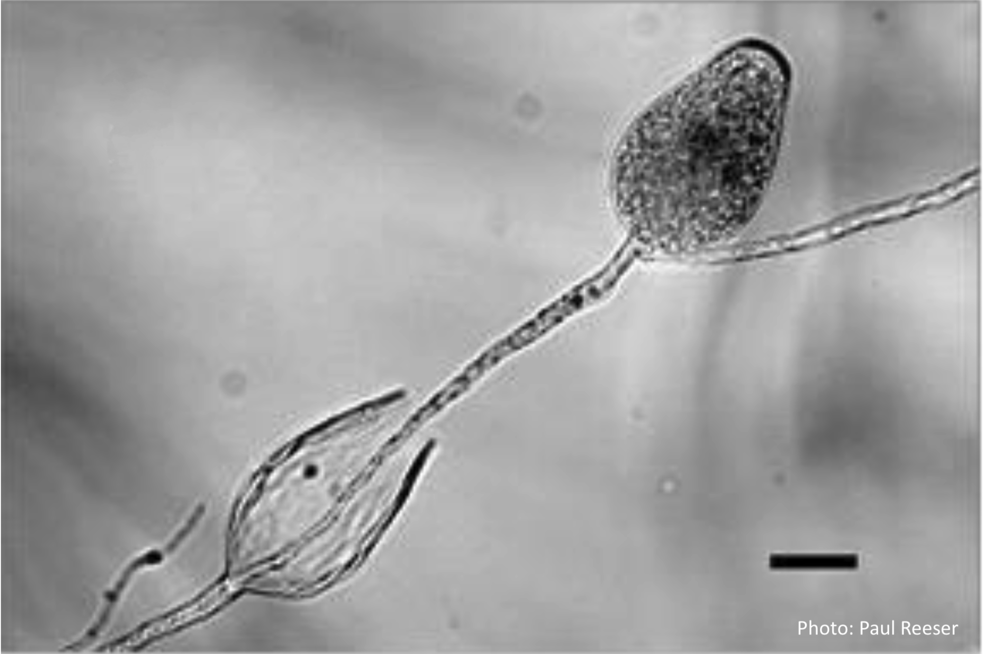 Phytophthora chlamydospora sporangia in water, showing subsporangial elongation. Bar is 20 µm. |