

Bole lesions in the tissues under the bark of a bleeding canker: distinct margin between healthy and disease tissues
Photo Gallery
|
P. siskiyouensis canker on Italian alder 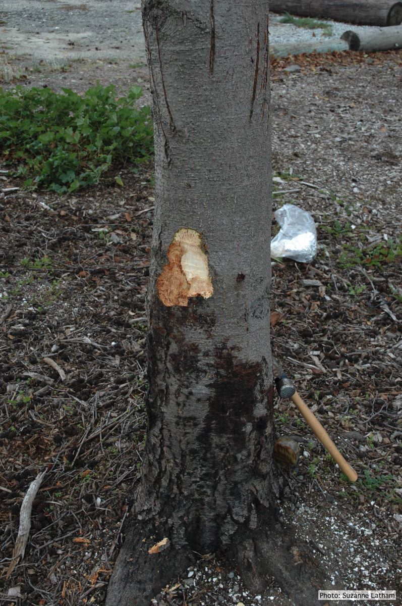 |
P. ramorum colony morphology on CMA PARP 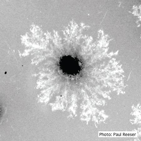 P. ramorum colony morphology on CMA PARP |
P. agathidicida oospores  Oospores of P. agathidicida in the roots of kauri seedlings inoculated with P. agathidicida. The root has been cleared with potassium hydroxide and bleached with peroxide, before being stained with Trypan Blue |
|
P. agathidicida oospores in planta  Oospores in the roots of kauri seedlings inoculated with P. agathidicida. The root has been cleared with potassium hydroxide and bleached with peroxide before being stained with trypan blue (scale bar =100 µm). |
P. chlamydospora sporangium 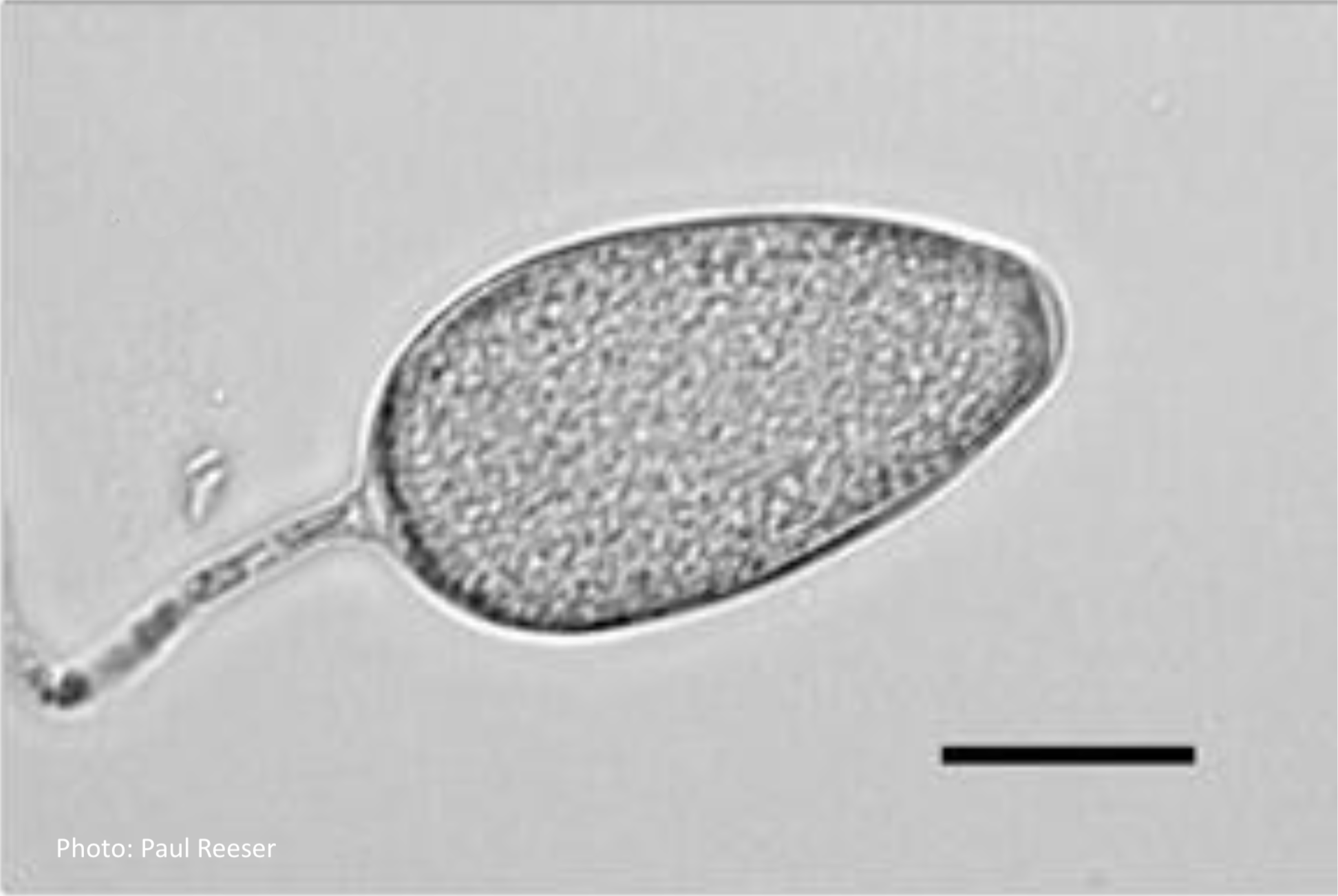 Phytophthora chlamydospora sporangium in water. Bar is 20µm. |
Austrocedrus associated with Mal del ciprés.  Austrocedrus with basal resin flow associated with Mal del ciprés. |
|
P. megakarya disease symptoms on Theobroma cacao fruit 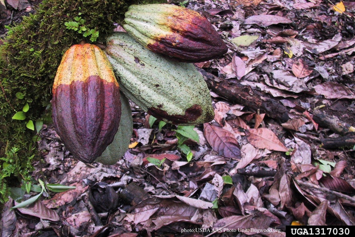 Disease symptoms on a cocoa pod |
P. agathidicia growth on PDA 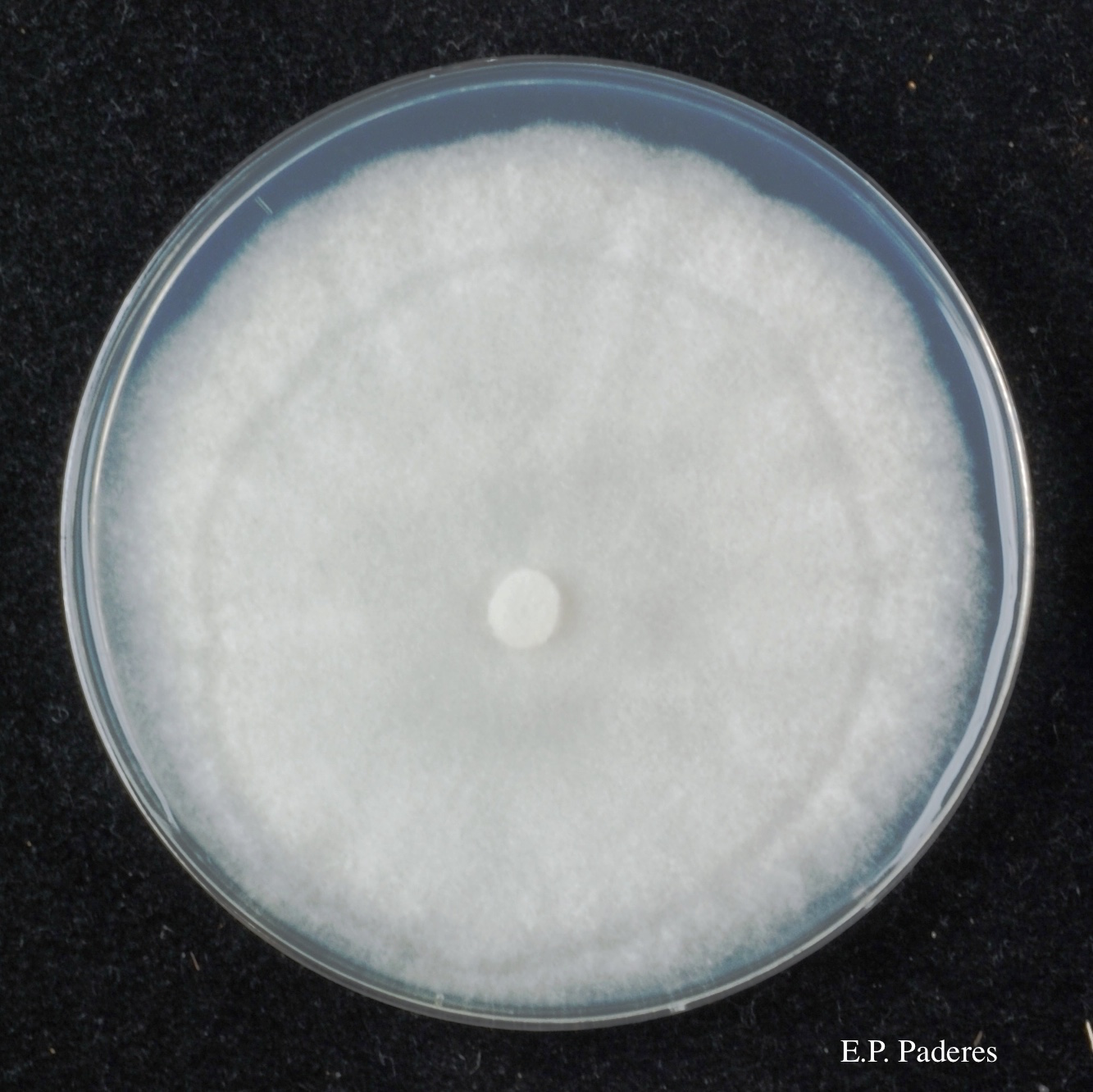 Colony morphology of ex-holotype ICMP 17027 after 10-days incubation at 20°C in the dark |
Basal canker on Port Orford Cedar stump  |
|
P. pluvialis hyphal swellings 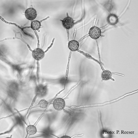 P. pluvialis hyphal swellings in water |
P. alni oogonia subspecies and variants 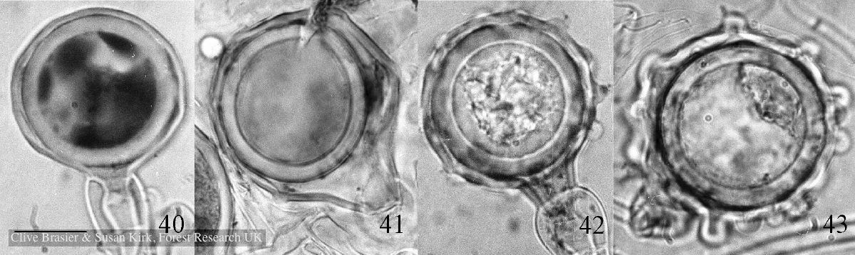 Fig. 40. P. alni subsp. uniformis. Fig. 41. P. alni subsp. multiformis German variant. Fig. 42. P. alni subsp. alni. Fig. 43. |
P. agathidicia disease symptoms on kauri 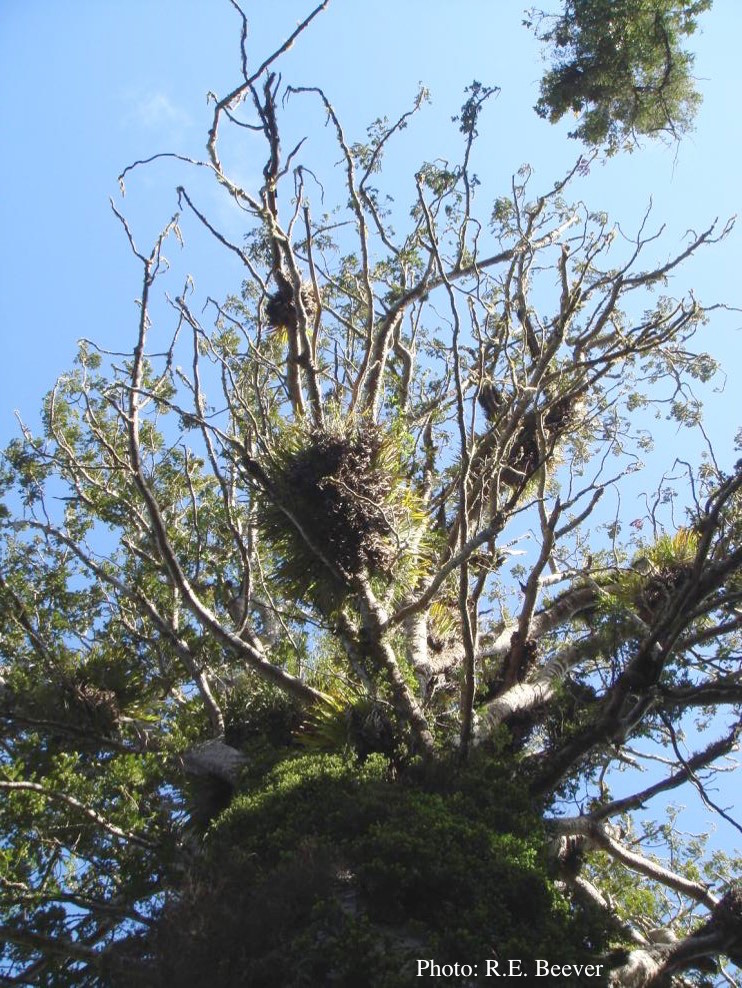 Crown decline of mature kauri, with branchlets with little or no leaves |