

Cottony colony morphology at 14 days at 20°C on PDA
Photo Gallery
|
P. cambivora colony morphology on PDA  |
P. megasperma colony morphology on V8  Colony morphology on V8 at 7 days |
P. palmivora sporangia, chlamydospores, hyphae 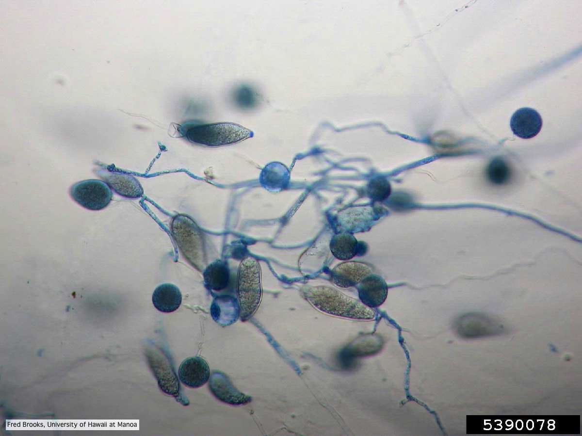 Sporangia (sporangiospores), chlamydospores, and hyphae stained with Cotton Blue |
|
P. nemorosa oogonium 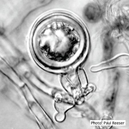 Oogonium with amphigynous antheridium |
P. frigida oogonium  Oogonium and oospore with amphigynous antheridium |
P. pseudotsugae sporangia  Broadly ovoid, papillate sporangia in water |
|
P. chlamydospora colony morphology on carrot agar 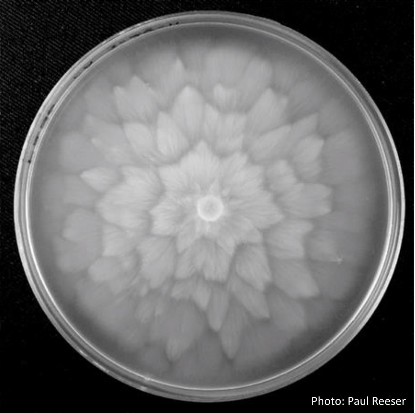 P. chlamydospora colony morphology on carrot agar |
P. chlamydospora chlamydospore 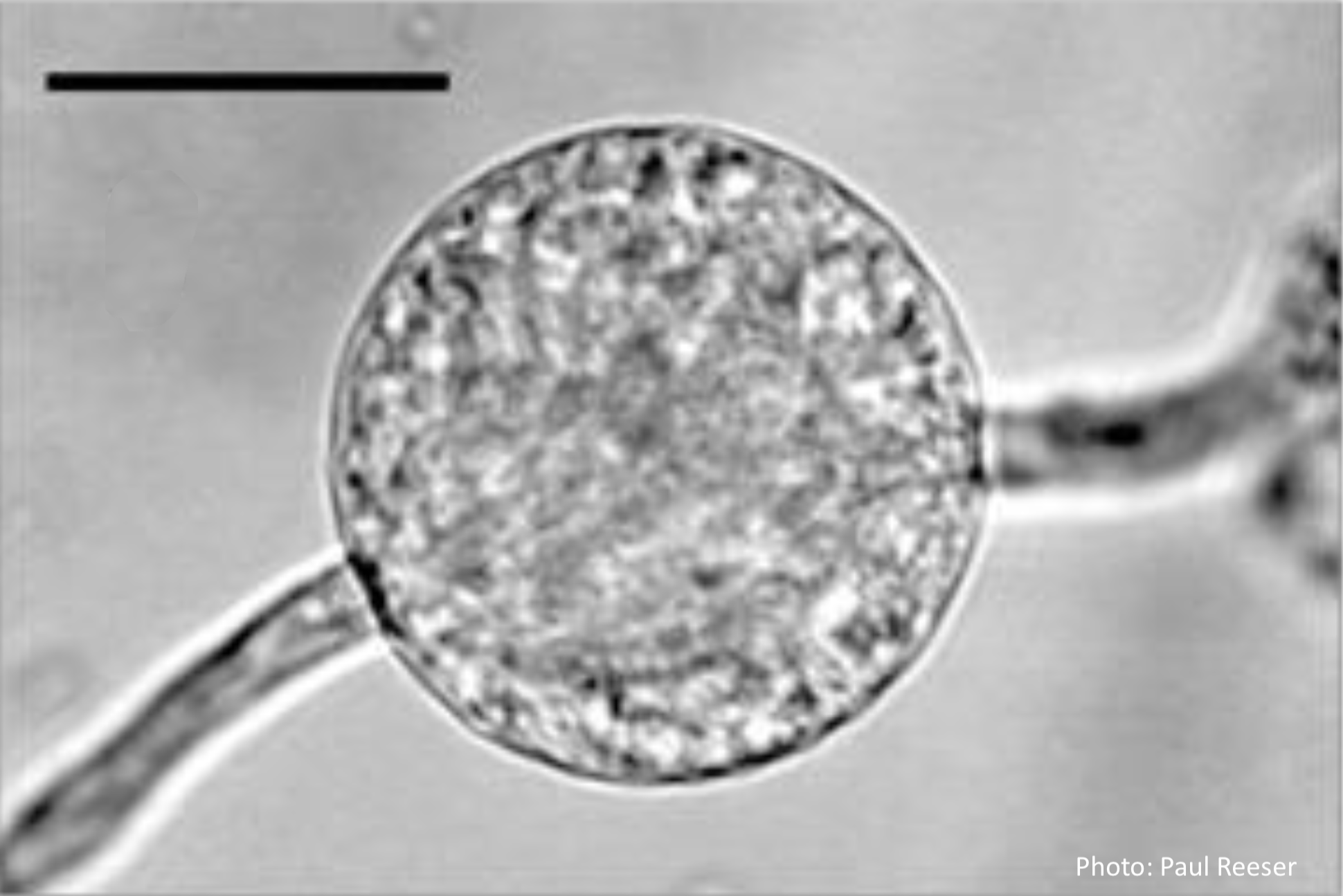 Phytophthora chlamydospora chlamydospore in agar. Bar is 20µm. |
P. tentaculata microscopic characteristics 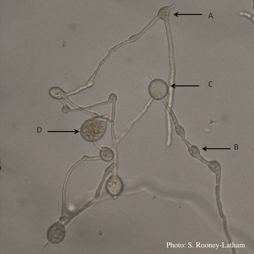 Hyphal swellings occuring at branching points of Mycelium (A), Intercalary hyphal swellings (B), Chlamydospore (C ), Sporangia (D) |
|
Sporangia showing ovoid and ovoid to spherical shape and papillate condition sporangia  Sporangia showing ovoid and ovoid to spherical shape and papillate condition |
Comparative gametangial morphology of Phytophthora Clade 5 species  Comparative gametangial morphology of Phytophthora Clade 5 species, with SEM (top) and light microscopy (bottom). P. heveae has smooth walled oogonia with funnel-shaped, amphigynous antheridia. P. agathidicida has mildly stipulate oogonia with globose amphigynous antheridia. P.cocois has mildly bullate oogonia with reflexed amphigynous antheridia. P. castaneae has coarsely bullate oogonium with rugose protuberances and narrow amphigynous antheridia (Weir et al. 2015). |
P. austrocedrae colony morphology on CMA 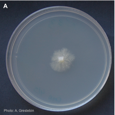 Colony morphology of P. austrocedrae at 16ºC after 4 weeks on CMA |