

P. pseudotsugae colony growth on V8 agar
Photo Gallery
|
P. pseudotsugae colony morphology on V8 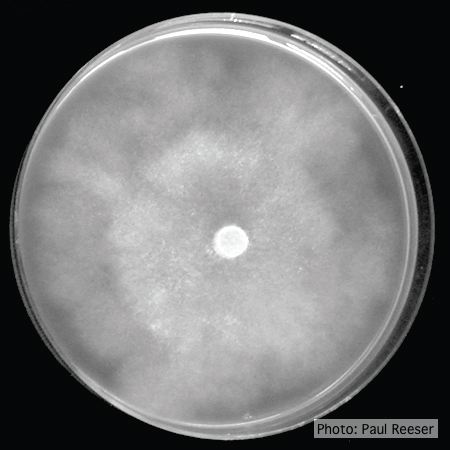 |
Dying Port Orford Cedar trees  |
P. cryptogea colony morpholgy on PDA 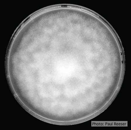 Colony morphology on PDA at 14 days |
|
P. kernoviae oogonia 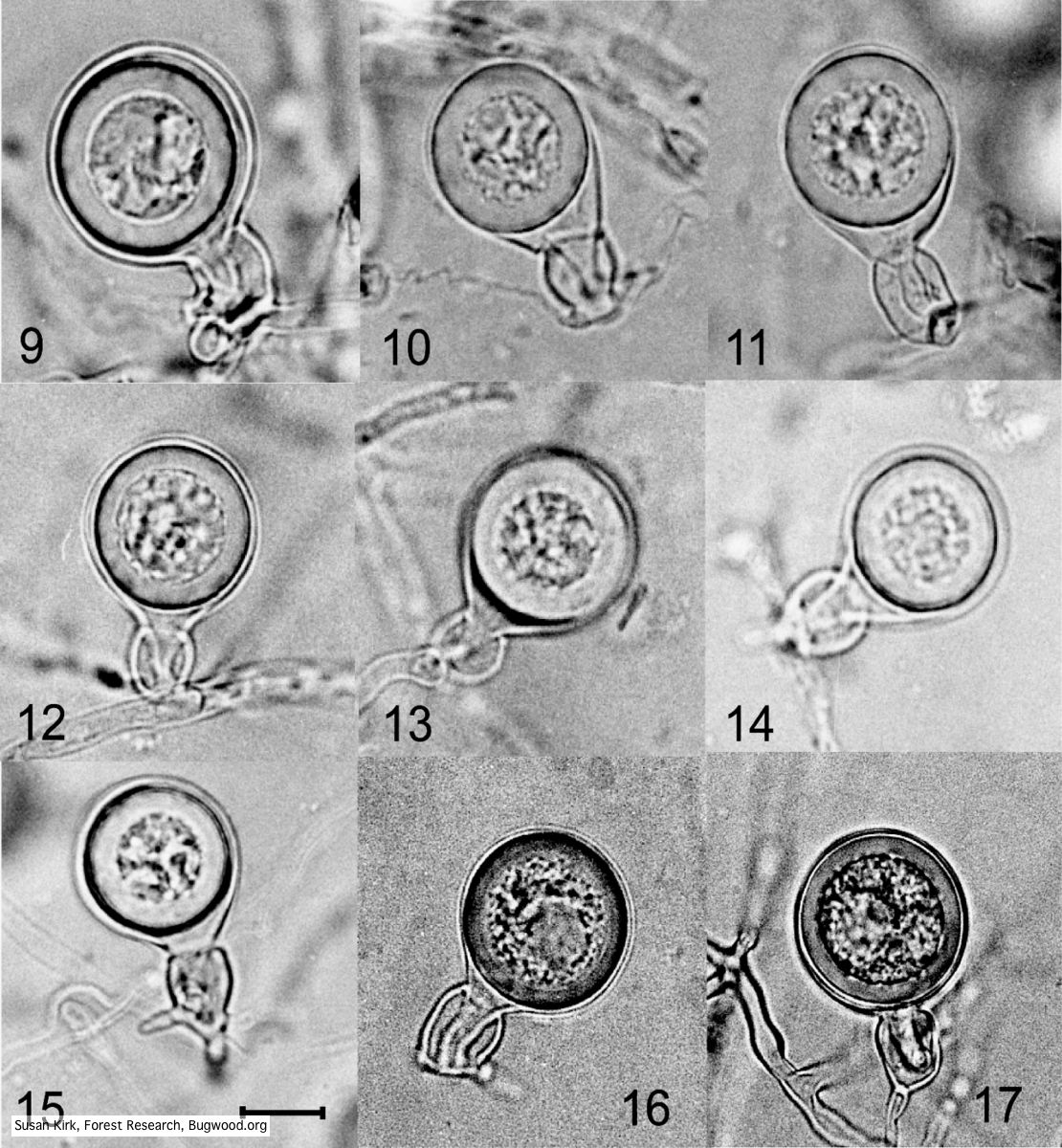 Mycol.Res 109, 853-859; Representative oogonia, antheridia and thick walled plerotic oospores of Phytophthora kernoviae. |
P. pluvialis sporangia.  P. pluvialis sporangia on tape peel from infected Douglas-fir needle. |
P. austrocedrae oogonia drawing  P. austrocedrae. Morphology of oogonia, oospores and antheridia. Bar: 10 mm. Greslebin et al. 2007 |
|
P. nicotianae colony morphology on PDA 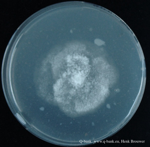 Phytophthora nicotianae CBS 321.49 PDA after 7 days at 24 degrees. Photo from Q-bank: www.q-bank.eu, Henk Brouwer (CBS-KNAW, Utrecht, The Netherlands) |
P. katsurae disease symptoms  Infected chestnut (Castanea) with bleeding canker |
P. pluvialis hyphal swellings 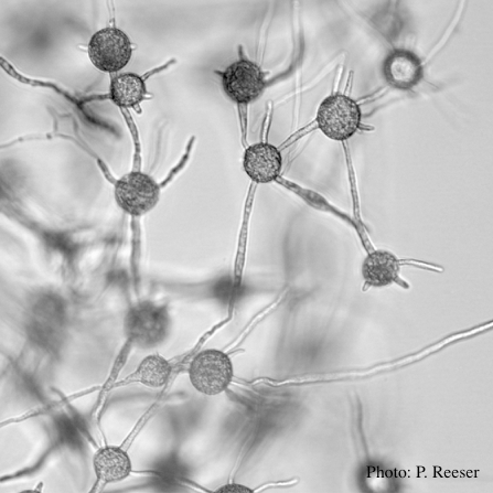 P. pluvialis hyphal swellings in water |
|
P. katsurae disease symptoms 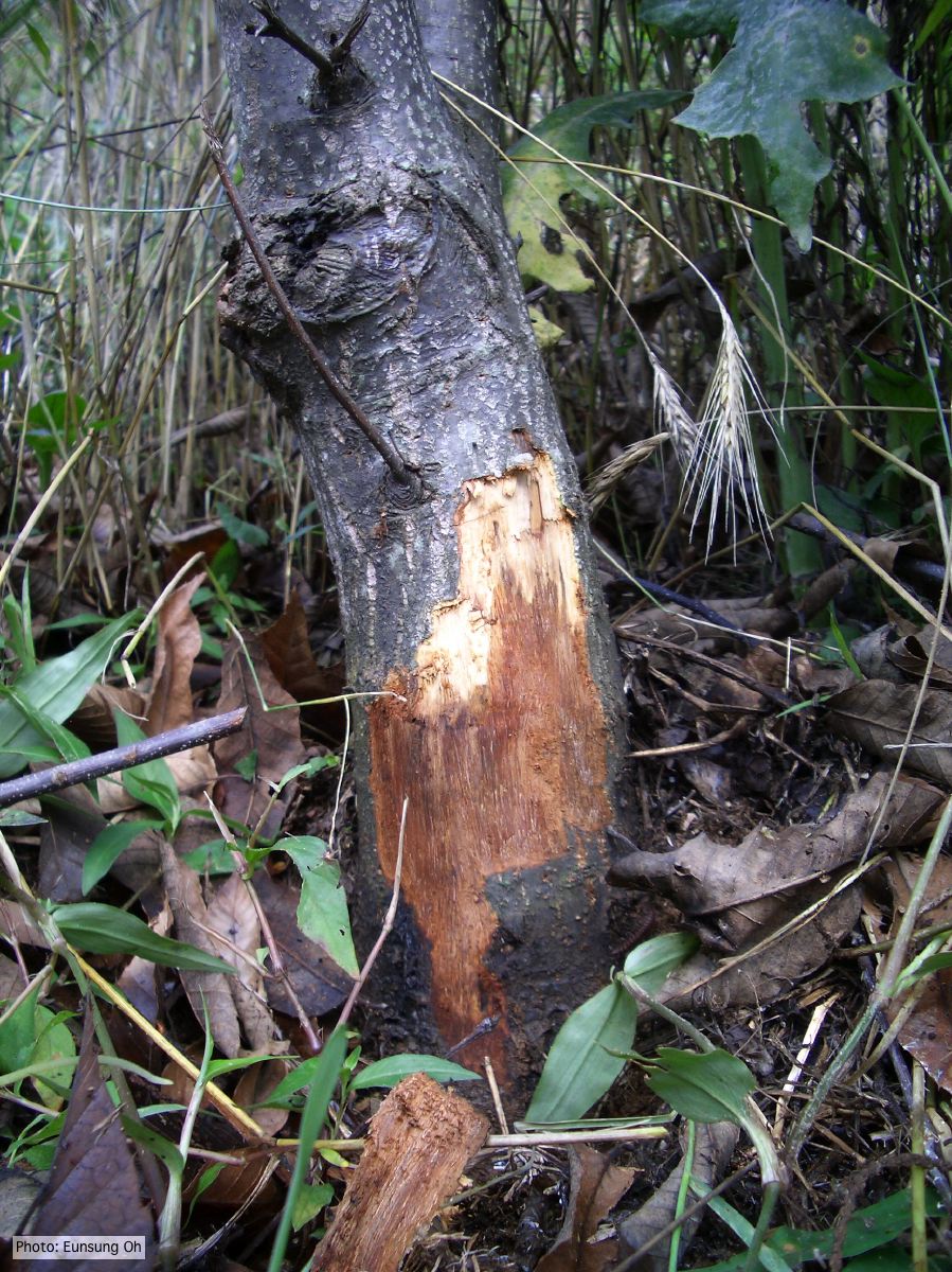 Infected chestnut tree with girdling canker on stem |
P. cactorum bleeding canker  Bleeding canker on European beech (Fagus sylvatica) |
P. megasperma oogonium 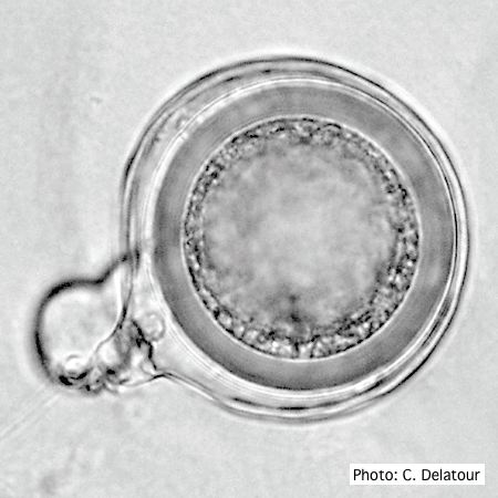 Oogonium with paragynous antheridia applied close to the ogonial stalk. |