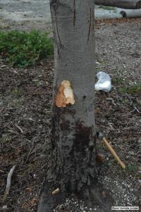
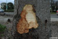
Bole lesions in the tissues under the bark of a bleeding canker: distinct margin between healthy and disease tissues
Photo Gallery
|
P. siskiyouensis canker on Italian alder |
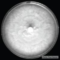
P. siskiyouensis colony morphology on V8 Colony morphology on V8 at 14 days |
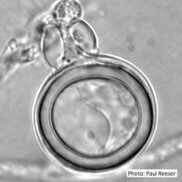
P. siskiyouensis oogonium with amphigynous antheridium P. siskiyouensis oogonium with amphigynous antheridium |
|
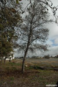
P. siskiyouensis disease symptoms on Italian alder Phytophthora collar rot on Italian alder trees: standing, dead tree |
P. siskiyouensis colony morphology on PDA Colony morphology on PDA at 14 days |
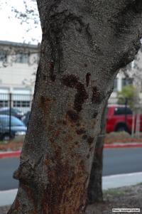
P. siskiyouensis canker on Italian alder Bleeding canker at the base of a tree and a sprinkler emitter (arrow) adjacent to the trunk |
|
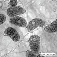
P. siskiyouensis sporangia Sporangia showing a variety of shapes and orientations of semi-papillae and sporangiophores |
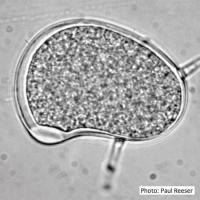
P. siskiyouensis sporangium P. siskiyouensis sporangium with lateral semi-papilla and subterminal, sub-basal insertion in the sporangiophore |
P. siskiyouensis canker on Italian alder Phytophthora collar rot on Italian alder trees: an isolated bleeding canker on the trunk |
|
P. siskiyouensis bleeding canker Close-up of margin area of bole lesions under the bark of a bleeding canker |
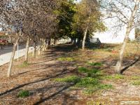
P. siskiyouensis disease symptoms on Italian alder Grove of dying trees in a commercial landscape in Foster City, CA |
P. siskiyouensis oogonium with paragynous antheridium P. siskiyouensis oogonium with paragynous antheridium |


