

Atypical decline caused by aerial infections in Scaër, France
Photo Gallery
|
P. lateralis on Port Orford cedar  |
P. kernoviae leaf wilt 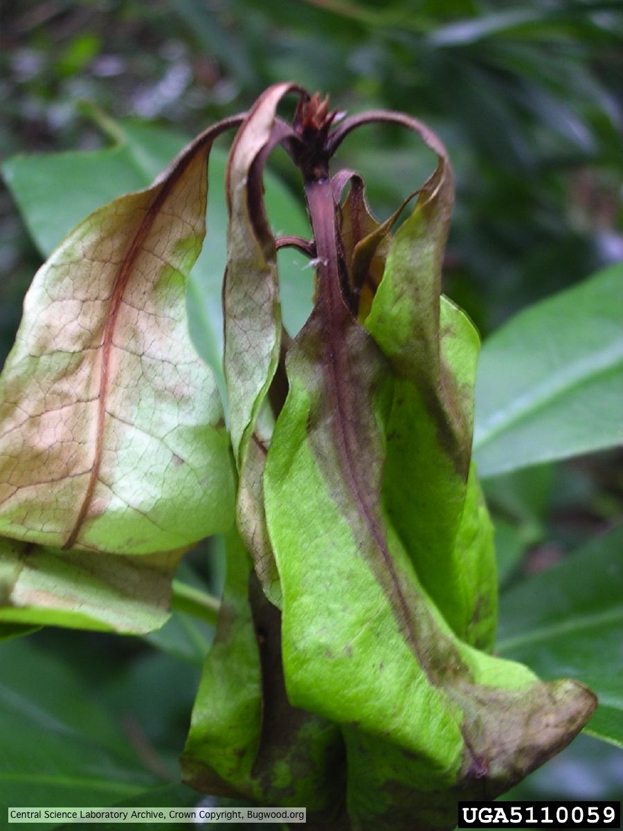 Necrosis of rhododendron leaves. |
P. cambivora colony morphology on MA 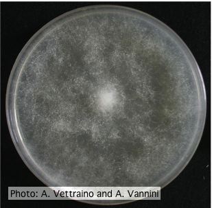 Uniform fluffy colony morphology at 14 days at 20°C on MA |
|
P. nicotianae sporangia  Noncaducous sporangium showing ovoid shape and papillate condition. (Fitopatol. bras. 2005) |
P. siskiyouensis sporangia 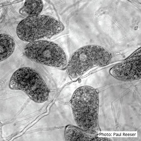 Sporangia showing a variety of shapes and orientations of semi-papillae and sporangiophores |
P. pinifolia on Pinus radiata 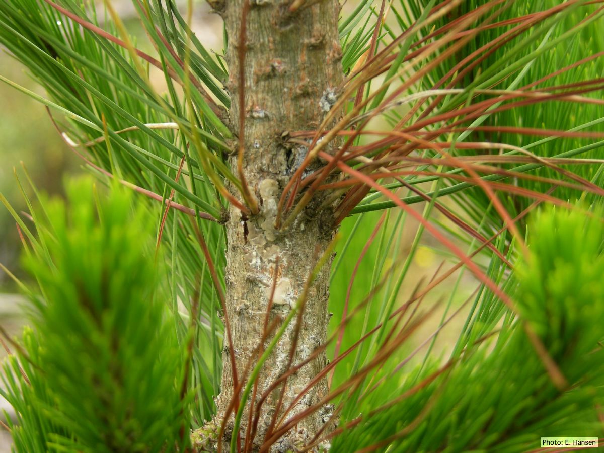 Pinus radiata, note Stem canker associated with necrotic needles. |
|
P. ramorum sporangia and chlamydospores 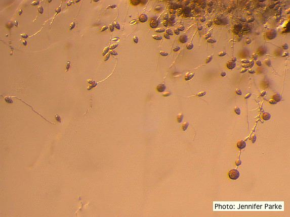 Sporangia and chlamydospores of P. ramorum |
P. pluvialis hyphal swellings 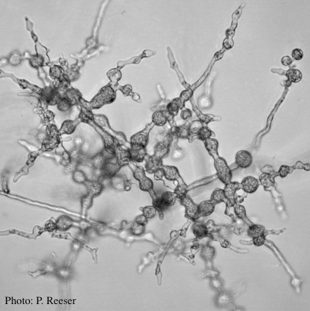 P. pluvialis hyphal swellings on agar |
P. austrocedrae hyphal swellings 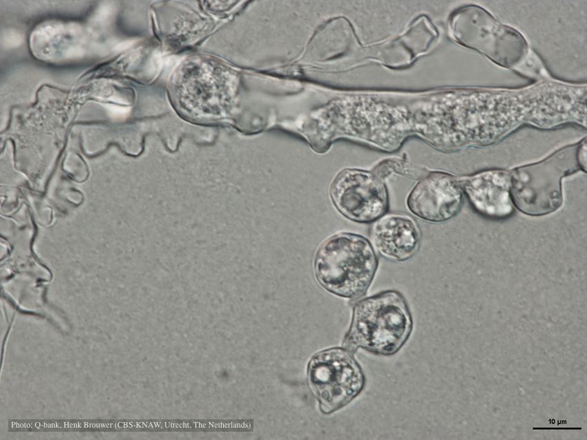 Hyphal swelling photo used with permission from Q-bank |
|
P. katsurae growth morphology on β-CMA 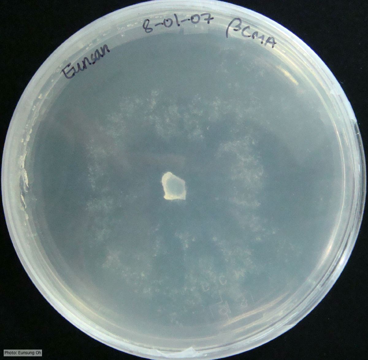 Growth morphology at 7 days on β-CMA |
P. palmivora sporangia 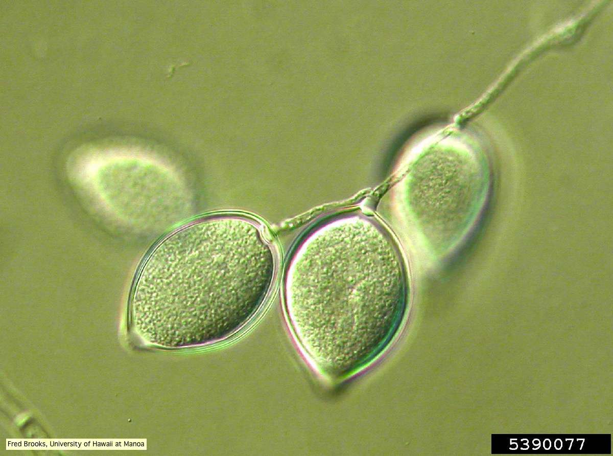 Sporangia (sporangiospores) showing sympodial branching |
P. megakarya sporangia (photo from Q-bank, used with permission). 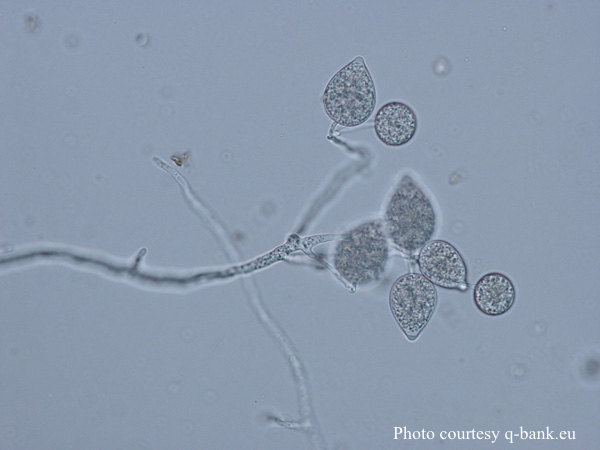 P. megakarya sporangia |