

Oogonium and oospore with amphigynous antheridium
Photo Gallery
|
P. frigida oogonium  |
P. alni in alder, Illwald, France  P. alni in alder, Illwald, France |
P. cambivora colony morphology on PDA 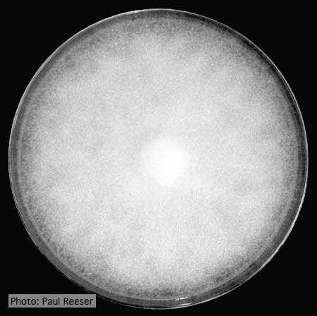 Colony morphology on PDA at 14 days |
|
P. kernoviae colony morphology on CMA PARPH 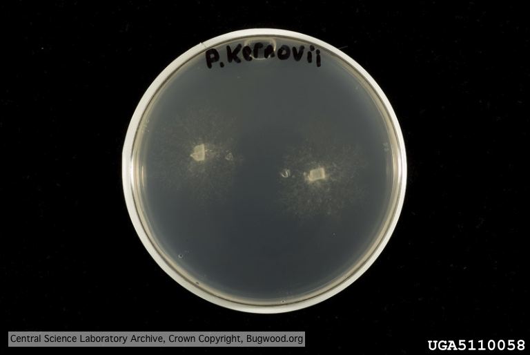 Organism grown on CMA PARP[H]; Plant disease 70, 1038-1043 |
P. pluvialis oogonium and antheridium  Oogonium and oospore with amphigynous antheridium |
P. lateralis on Port Orford cedar  Lesion caused by aerial infection on Chaemacyparis lawsoniana in Lopérec, France |
|
P. pluvialis symptoms on Douglas-fir  Red needle cast symptoms on Douglas-fir in western Oregon, 2015 |
P. alni sporangia 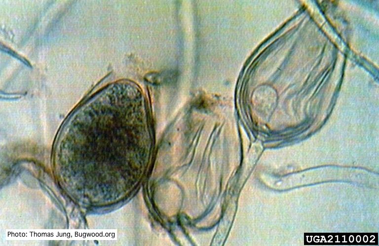 Non-papillate sporangia of P. alni showing nested proliferation. |
P. pinifolia on Pinus radiata 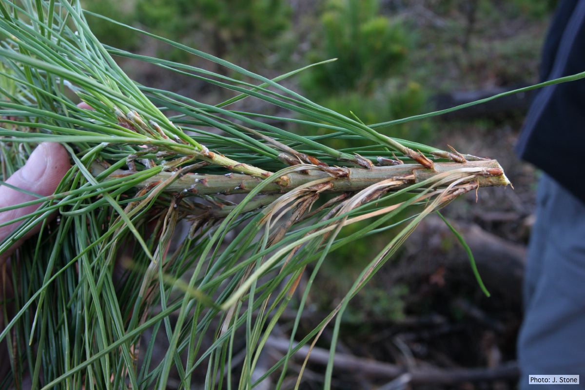 Pinus radiata, note grey and collapsed needle bases |
|
P. cinnamomi on Banksia  Death of woodland Banksia, Western Australia |
P. kernoviae canker  Bole lesion on Fagus sylvatica |
Mal del ciprés, dead and dying trees along river 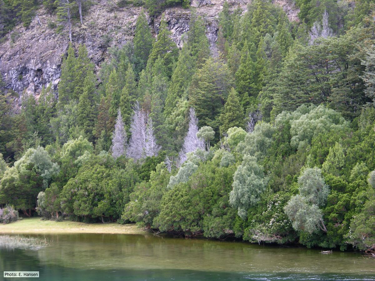 Mal del ciprés, dead and dying trees along river |