

Sporangium with internal proliferation, photo from Q-bank, used with permission.
Photo Gallery
Site will be retired 9/1/2026
This site is no longer being developed and will be retired on September 1, 2026. Please contact us if you have any questions or would like to provide support to continue the project.
|
P. pinifolia sporangia 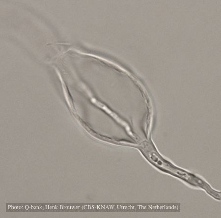 |
P. ramorum zoospores 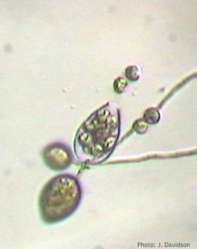 Sporangium of P. ramorum releasing zoospores |
P. tentaculata on V-8 media 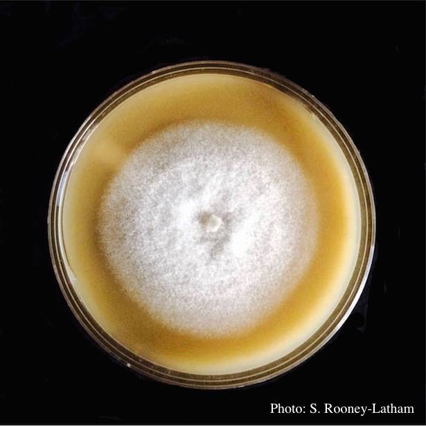 Culture of P. tentaculata on V-8 media |
|
Stain from Port Orford Cedar root disease  Stain from Chamaecyparis lawsoniana root disease on the Smith River |
P. pseudotsugae sporangium 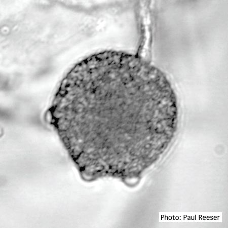 Broadly ovoid, papillate sporangium in water |
P. pluvialis on Pinus radiata in New Zealand 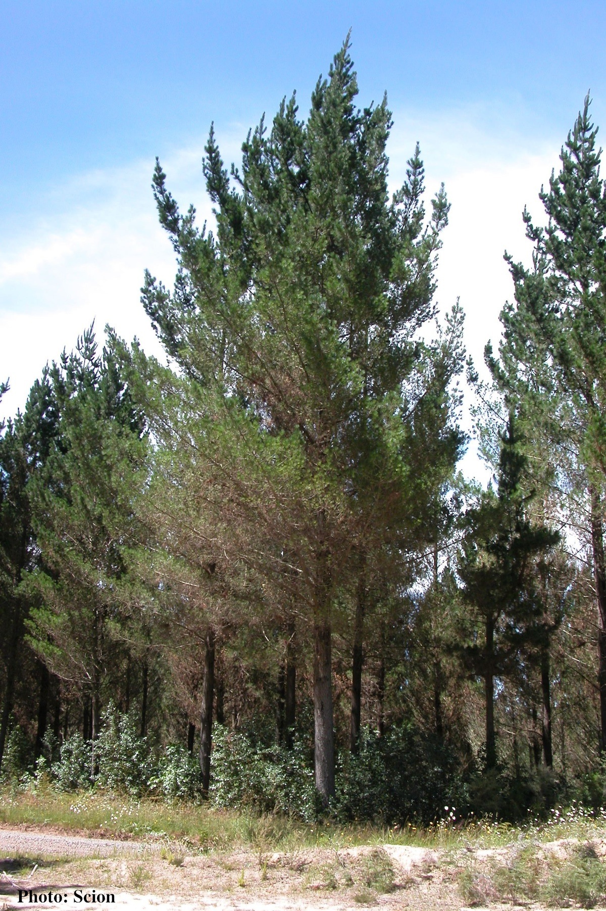 A stand of Pinus radiata trees affected by red needle cast disease. Note that frequently only the lower part of the crown is affected. |
|
P. palmivora sporangia, chlamydospores, hyphae 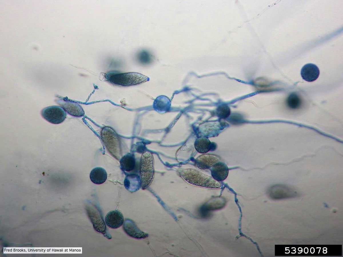 Sporangia (sporangiospores), chlamydospores, and hyphae stained with Cotton Blue |
P. ramorum canker 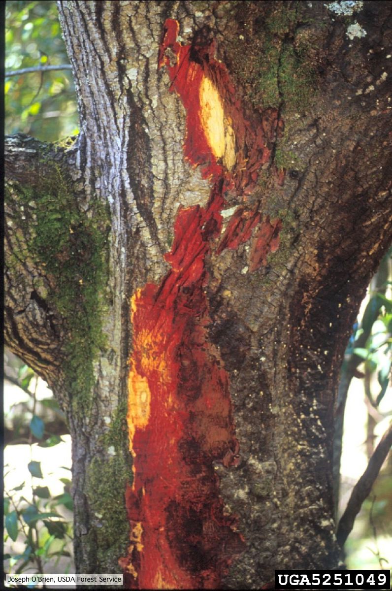 Bark discoloration and zone lines in coast live oak (Quercus agrifolia) |
P. tentaculata disease symptoms on California mugwort 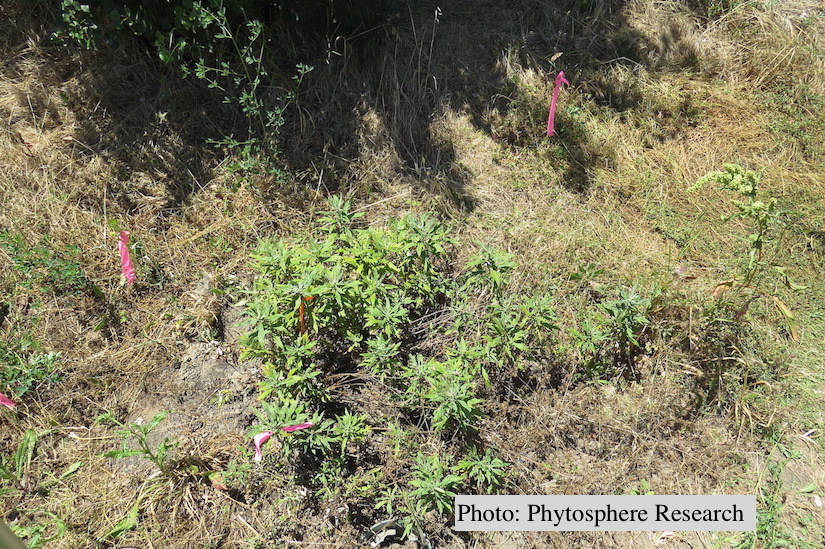 Outplanted California mugwort (Artemisia douglasiana) infected with P. tentaculata, 4.5 years after planting. Plant shows stunting and chlorosis. (P. cryptogea and P. lacustris were also baited from roots/soil of this plant). |
|
P. palmivora chlamydospore 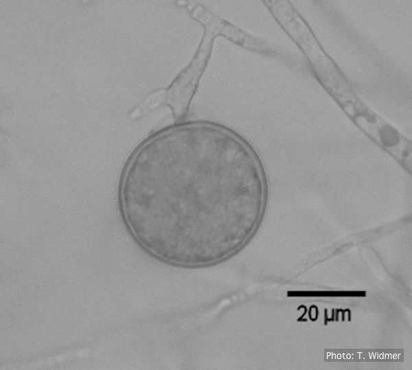 Terminal chlamydospore of P. palmivora |
P. frigida symptoms 2 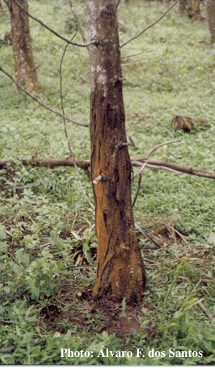 Symptoms of gummosis on black wattle |
P. alni subsp alni sporangium 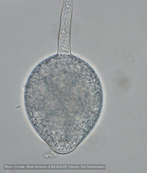 Non-papillate, non caducous sporangium, photo used with permission from Q-bank |