
Phytophthora pluvialis
Site will be retired 9/1/2026
This site is no longer being developed and will be retired on September 1, 2026. Please contact us if you have any questions or would like to provide support to continue the project.
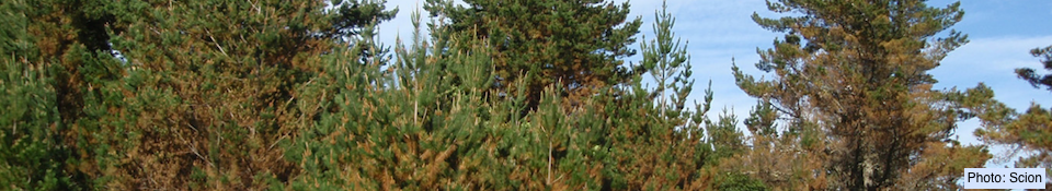
Phytophthora pluvialis Reeser, Sutton, and E. Hansen was first discovered in streams, soil, and raintraps in Curry County, Oregon, USA, in areas dominated by native forest of mixed tanoak and Douglas-fir (Reeser et al., 2013). On rare occasions it was isolated from lesions on tanoak bark or twigs, but was only weakly pathogenic when inoculated into tanoak stems. In New Zealand, Phytophthora pluvialis was found to cause red needle cast on radiata pine (Dick et al., 2014). In Oregon the species is now associated with Douglas-fir, and experimentally has been shown to cause needle cast and twig cankers on seedling trees, both artificially inoculated and exposed beneath the canopy of Douglas-fir plantations (Hansen et al., 2014). The pathogen has not been associated with canker formation or infection of the harvested stem of radiata pine (Hood et al., 2014).
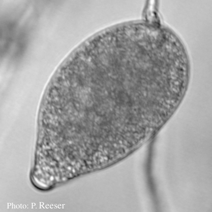
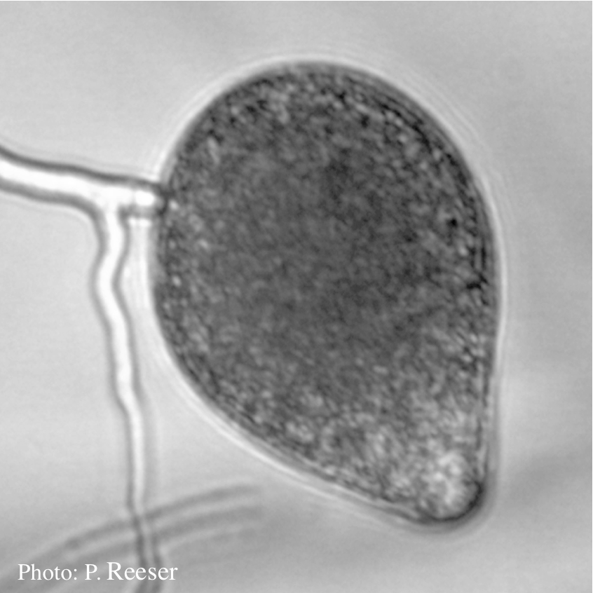
Sporangia showing typical ovoid shape and semi-papillate condition.
Sporangia (average 53–67 μm by 39–45 μm) formed in water, ovoid or slightly irregular (occasionally bifurcate), semi-papillate, terminal or sub-terminal on simple or sympodial sporangiophores. Sporangia are partially caducous with variable, medium length (5–22 μm) pedicels. Hyphal swellings formed in water are globose and intercalary with radiating hyphae forming loose networks. Hyphal swellings formed in agar are variable. Oogonia (average 27 μm to 34 μm), formed in single agar culture, globose, smooth, terminal with amphigynous antheridia. Oospores (average ca. 27–29 μm), globose, aplerotic.
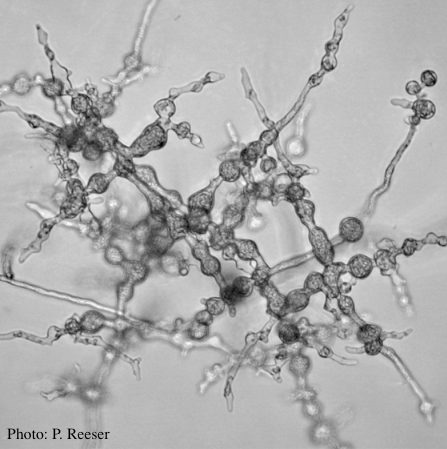

Left: P. pluvialis hyphal swellings on agar. Right: Oogonia and oospores with amphigynous antheridia.
Phytophthora pluvialis resides in ITS Clade 3 (Cooke et al., 2000) along with P. ilicis Buddenhagen & Young, P. nemorosa E.M. Hansen & Reeser, P. pseudosyringae T. Jung & Delatour, and P. psychrophila T. Jung & E.M. Hansen. It is readily distinguished by ITS DNA sequence from related species in Clade 3.
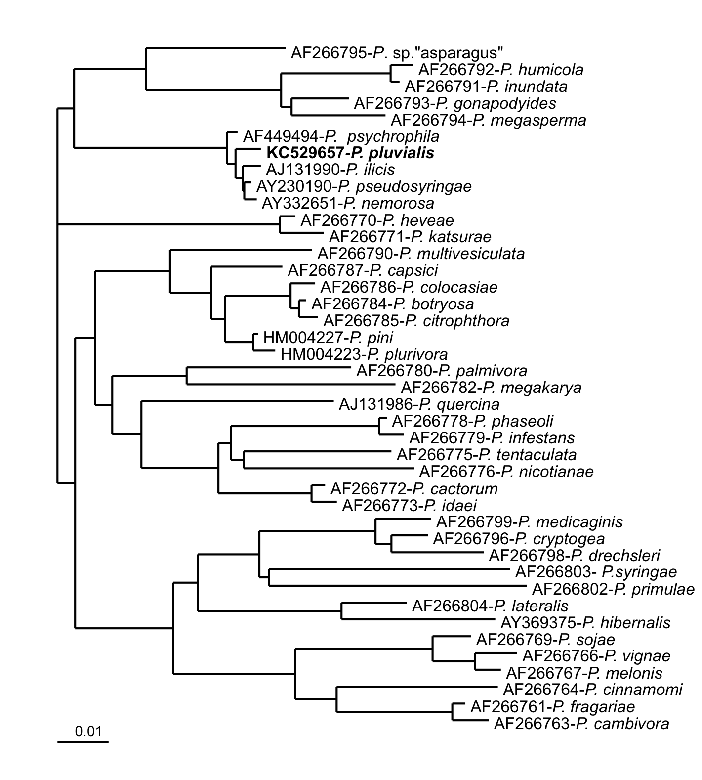
Phylogenetic relations based on rDNA-ITS sequences (Reeser et al., 2013).
The colony pattern on carrot agar is angular and petaloid, with hyphae appressed. Radial growth rate on carrot agar is about 1.2–1.4 mm/d at 15°C. Temperature optimum was ca. 15°C, min. < 5°C, max. ca. 25°C.


Colony morphology of two different isolates on carrot agar at 20 days.
P. pluvialis is most readily recognized by its semi-papillate sporangia, amphigynous antheridia, and distinctive hyphal swellings formed both in agar and in water. It is most similar to P. nemorosa, but growth rate of P. pluvialis at optimum temperature is much slower.
For more information about Phytophthora pluvialis, visit our Disease, Education and Management materials, and Reference sections.