

Noncaducous sporangium showing ovoid shape and papillate condition. (Fitopatol. bras. 2005)
Photo Gallery
|
P. nicotianae sporangia  |
P. cambivora bole canker 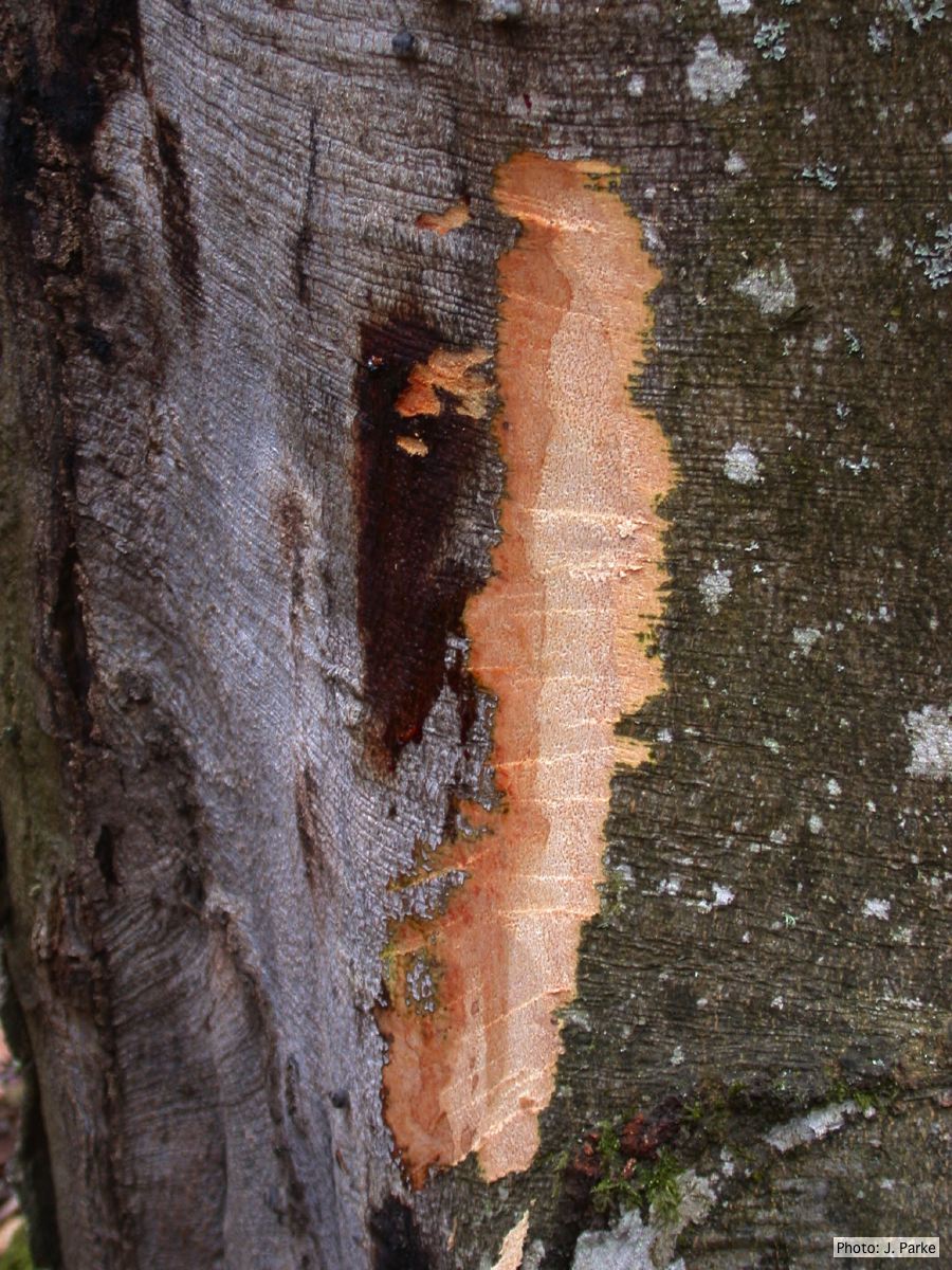 Fagus sylvatica bole canker |
P. cambivora colony morphology on MA 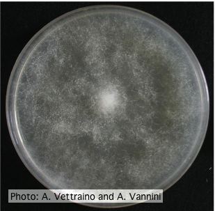 Uniform fluffy colony morphology at 14 days at 20°C on MA |
|
P. pluvialis symptoms on Douglas-fir needles  Symptoms of red needle cast on Douglas-fir needles |
Black pod disease of cacao caused by P. palmivora 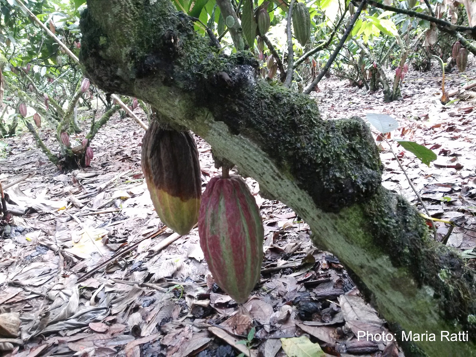 Black pod of cacao in Ecuador caused by P. palmivora (see lesioned fruit on left). |
P. alni oogonia subspecies and variants 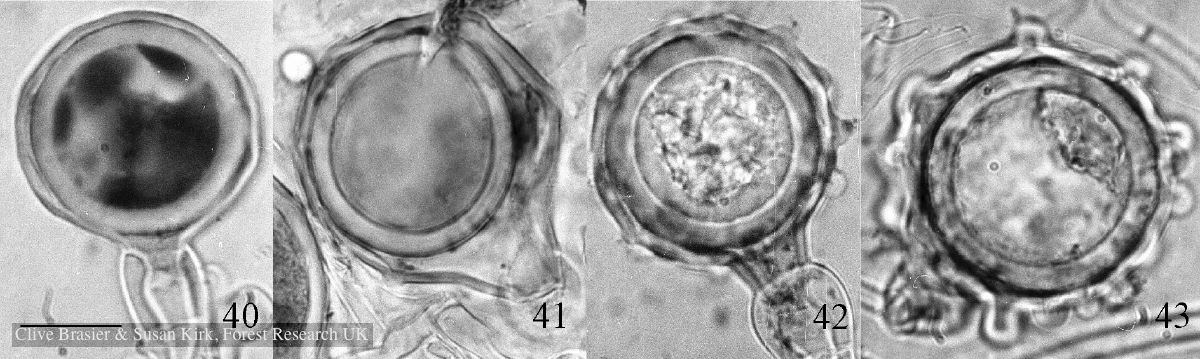 Fig. 40. P. alni subsp. uniformis. Fig. 41. P. alni subsp. multiformis German variant. Fig. 42. P. alni subsp. alni. Fig. 43. |
|
P. austrocedrae - Mal del ciprés in Argentina 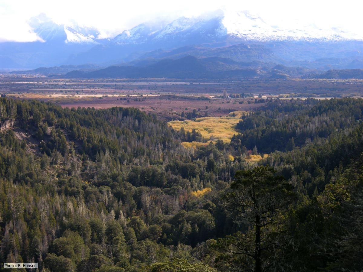 Mal del ciprés looking toward Rio Grande, Chubut Province, Argentina |
Vehicle washing  Truck washing to avoid spread of P. lateralis |
P. kernoviae leaf wilt 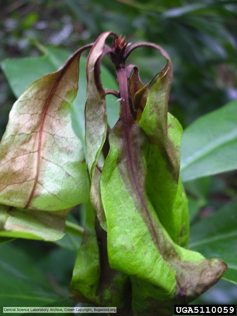 Necrosis of rhododendron leaves. |
|
P. boehmeriae oogonia  Oogonia and oospores with amphigynous antheridia |
P. lateralis on Port Orford cedar  Atypical decline caused by aerial infections in Scaër, France |
P. megakarya oospore
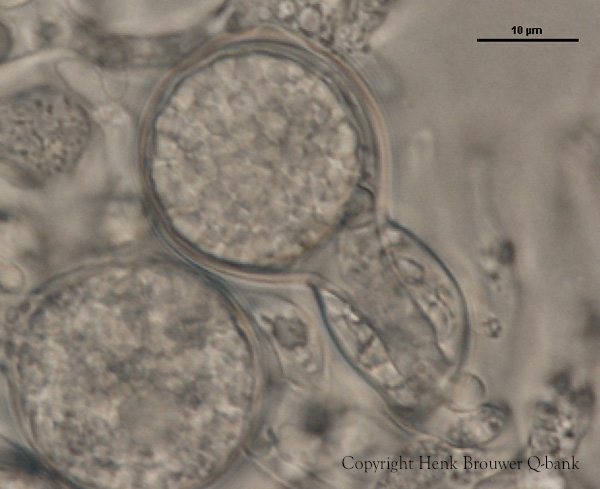
P. megakarya oogonia, oospore, and antheridium
|