

Mal del ciprés, dead and dying trees along river
Photo Gallery
Site will be retired 9/1/2026
This site is no longer being developed and will be retired on September 1, 2026. Please contact us if you have any questions or would like to provide support to continue the project.
|
Mal del ciprés, dead and dying trees along river 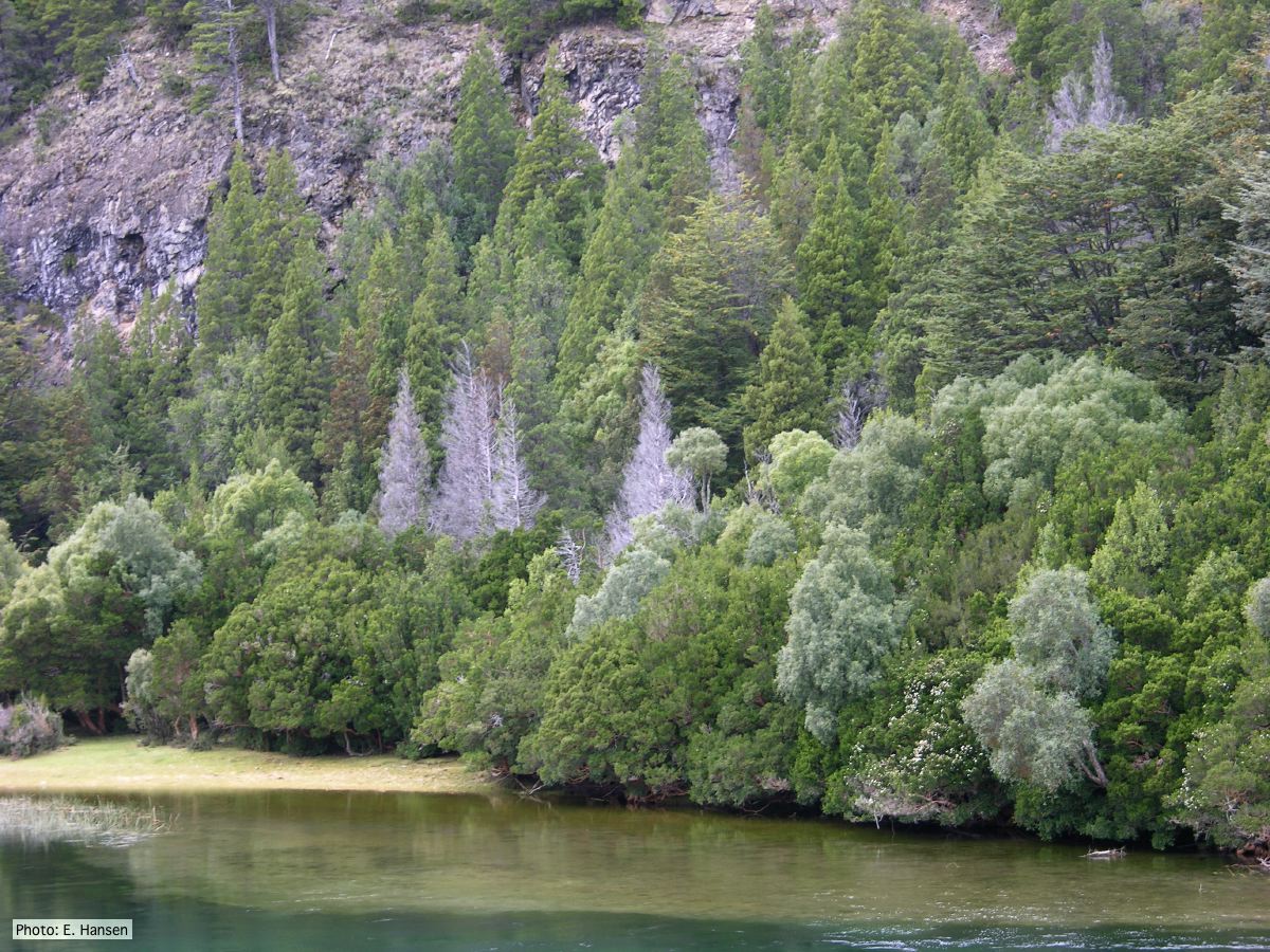 |
P. lateralis on Port Orford cedar 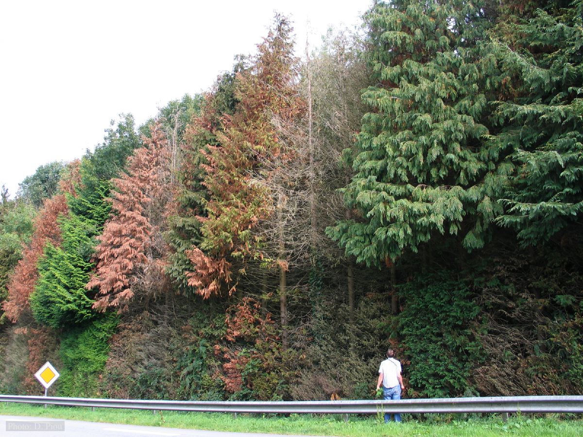 Typical decline of Chaemacyparis lawsoniana in Landrévarzec, France. |
P. alni in riparian alder, Scotland 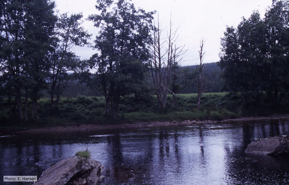 P. alni in riparian alder, Scotland |
|
Necrotic lesion in phloem caused by P. austrocedrae  Necrotic lesion in phloem with resin pocket caused by P. austrocedrae |
Dead Port Orford Cedar 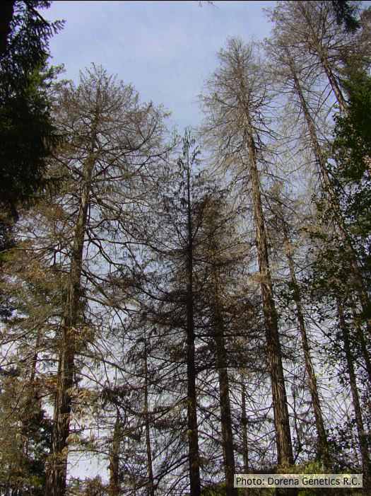 Dead Chamaecyparis lawsoniana, BLM Roseburg District in Oregon |
P. cambivora colony morphology on MA 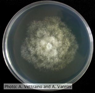 Rosacous colony morphology at 14 days at 20°C on MA |
|
P. pluvialis on Pinus radiata needle  Clusters of sporangia emerge from stomata of an infected radiata pine needle. |
P. megasperma colony morphology on PDA  Colony morphology on PDA at 7 days |
P. cactorum colony morphology on V8  Colony morphology on V8 at 14 days |
|
P. austrocedrae necrotic lesion in phloem 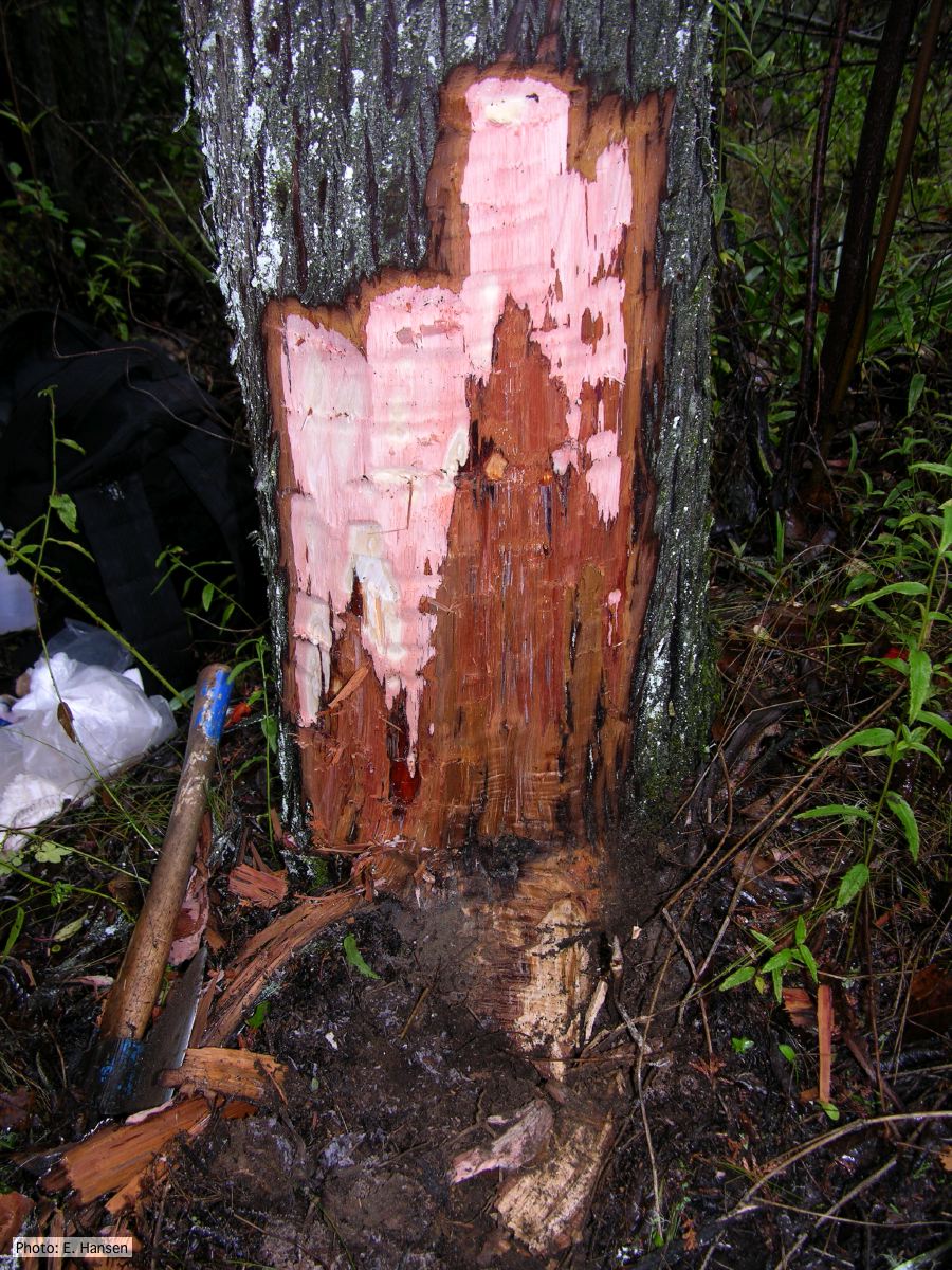 P. austrocedrae - necrotic lesion in phloem |
P. cactorum oogonium 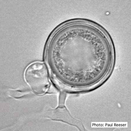 Oogonium with paragynous antheridia close to oogonial stalk. Oospores are slightly aplerotic. |
P. austrocedrae colony morphology on Tomato juice agar 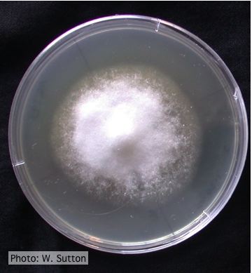 Colony morphology of P. austrocedrae at 16 ºC after 4 weeks on Tomato juice agar |