

Dead Banksia sp. in a Kwongan heathland on mineral sand near Eneabba, Western Australia recently killed by root and collar rot caused by Phytophthora arenaria
Photo Gallery
Site will be retired 9/1/2026
This site is no longer being developed and will be retired on September 1, 2026. Please contact us if you have any questions or would like to provide support to continue the project.
|
P. arenaria disease symptoms on Banksia 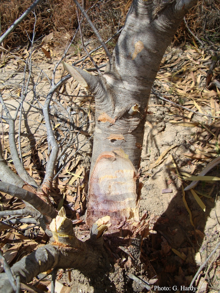 |
P. austrocedrae hyphal swellings 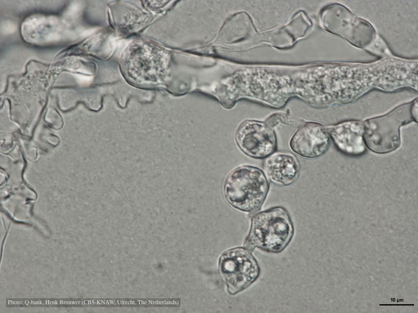 Hyphal swelling photo used with permission from Q-bank |
P. austrocedrae colony morphology on Tomato juice agar 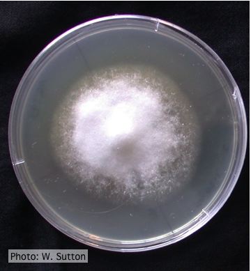 Colony morphology of P. austrocedrae at 16 ºC after 4 weeks on Tomato juice agar |
|
Mat to control spread of P. agathidicida 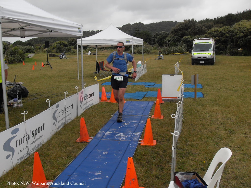 Jogger running over a plastic-reinforced, foam-mat containing a 2% solution of Trigene™ Advance (quaternary ammonium compound) as part of a cross-country event, in the Waitakere Regional Park |
P. kernoviae leaf wilt 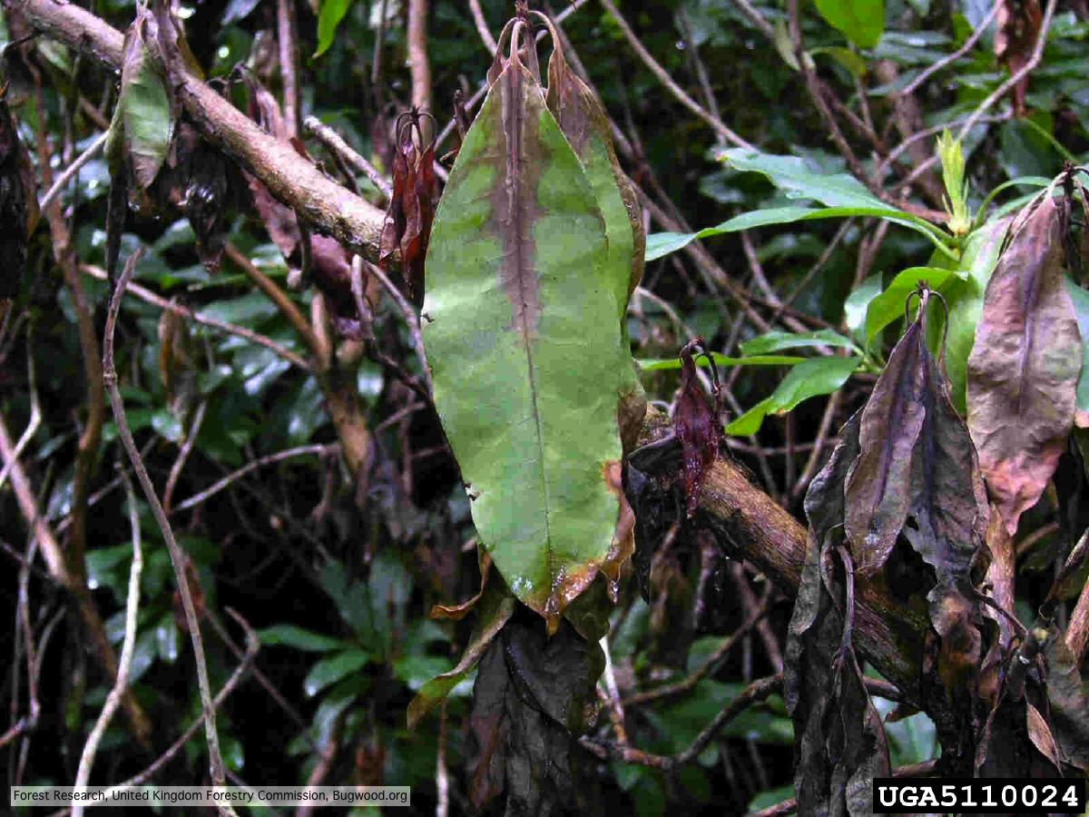 Wilted leaf of infected rhododendron |
Stain from Port Orford Cedar root disease  Stain from Chamaecyparis lawsoniana root disease on the Smith River |
|
P. pluvialis symptoms on Douglas-fir 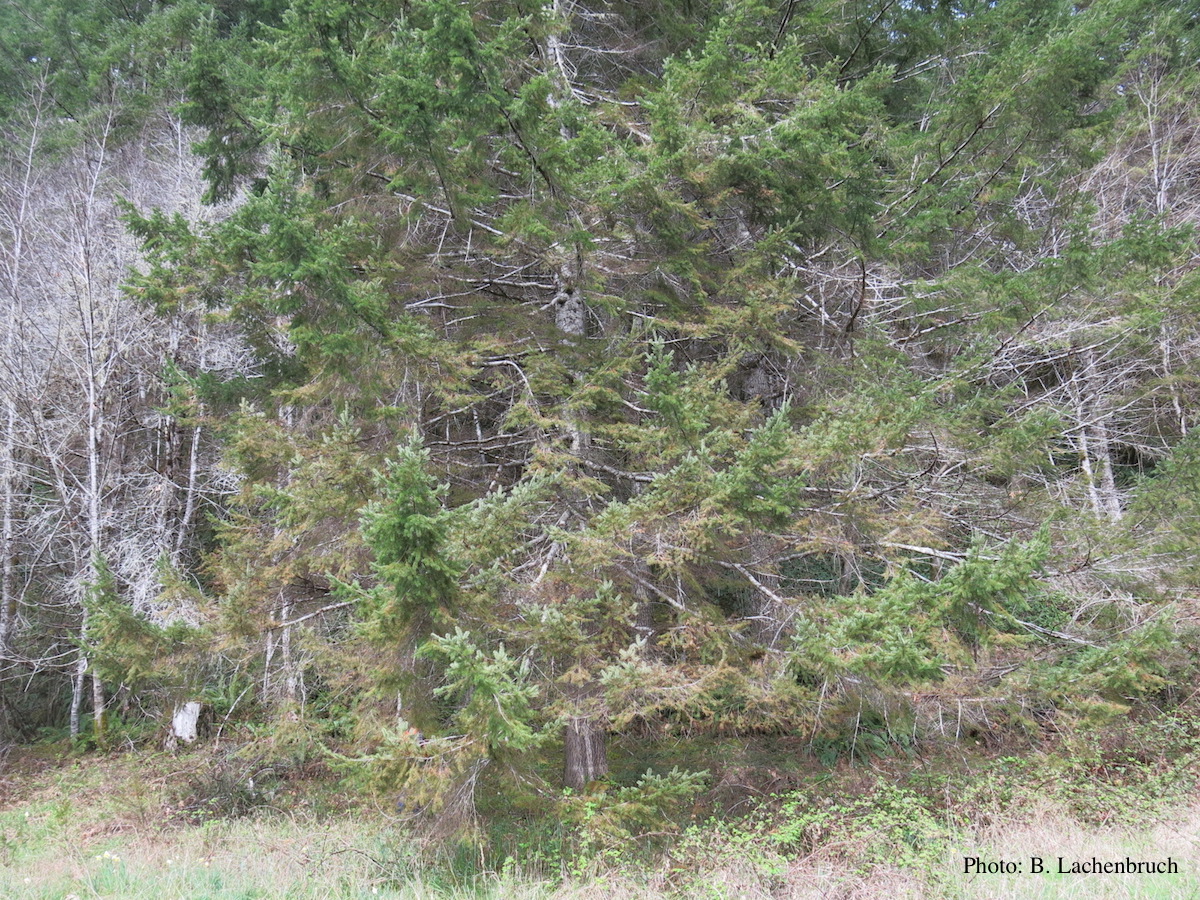 Red needle cast symptoms on Douglas-fir in western Oregon, 2015 |
P. lateralis on Port Orford cedar 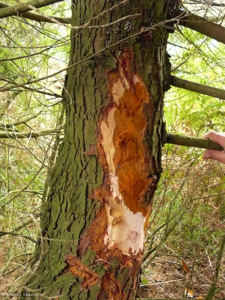 Bole lesion on Chaemacyparis lawsoniana in Lopérec, France |
P. pseudosyringae sporangium 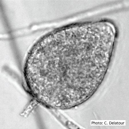 Ovoid, semipapillate sporangia showing medium length pedicel |
|
P. cambivora oogonium  Bullate oogonium and and two-celled amphigynous antheridium |
P. palmivora sporangia 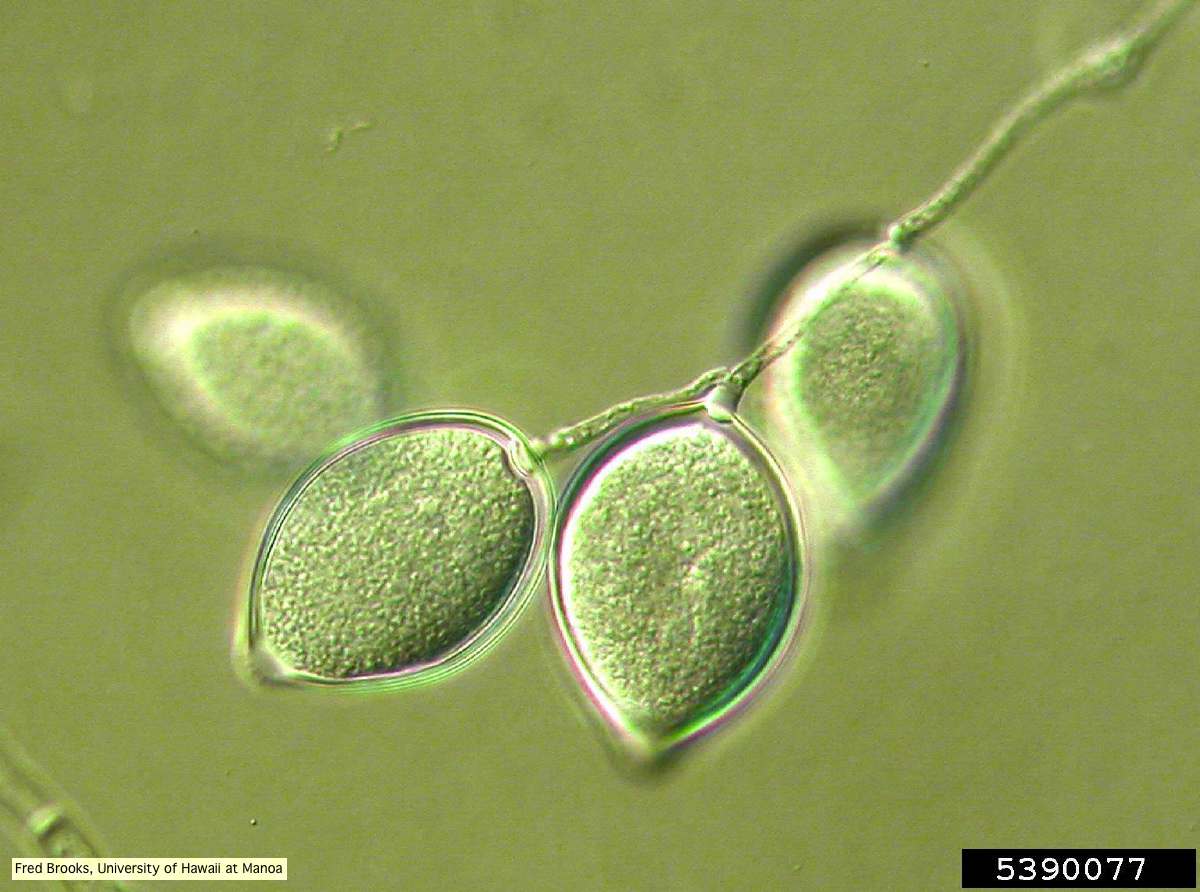 Sporangia (sporangiospores) showing sympodial branching |
P. agathidicida sporangium 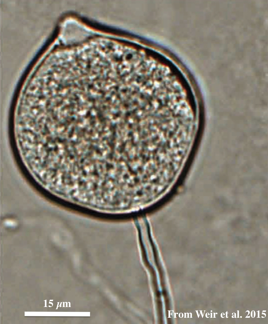 Globose to ovoid-ellipsoid, papillate sporangium |