

Smooth-walled oogonium of P. alni (Swedish variant) with oospore and amphigynous antheridium.
Photo Gallery
Site will be retired 9/1/2026
This site is no longer being developed and will be retired on September 1, 2026. Please contact us if you have any questions or would like to provide support to continue the project.
|
P. alni oogonia 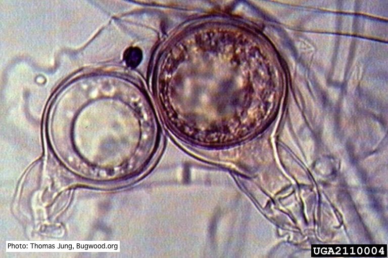 |
Growth of P. megakarya on PDA 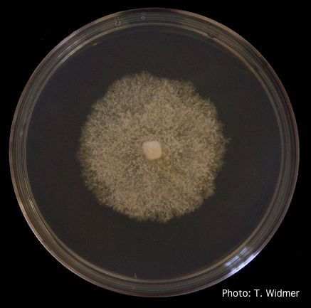 Growth of P. megakarya on potato dextrose agar |
Dead and healthy Port-Orford cedar seedlings  Port-Orford-cedar seedlings planted to test for Phytophthora lateralis resistance at the Dorena Genetic Resource Center |
|
Mal del ciprés, dead and dying trees along river 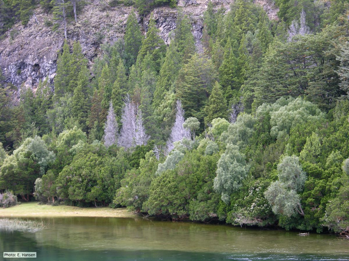 Mal del ciprés, dead and dying trees along river |
P. pluvialis symptoms on Douglas-fir 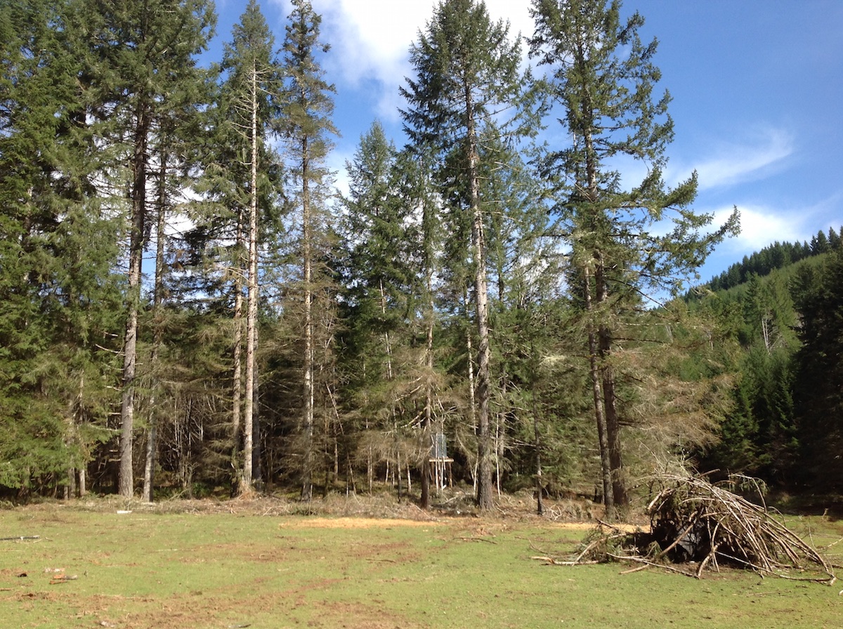 P. pluvialis symptoms of red needle cast on Douglas-fir, western Oregon 2015 |
P. frigida symptoms 3 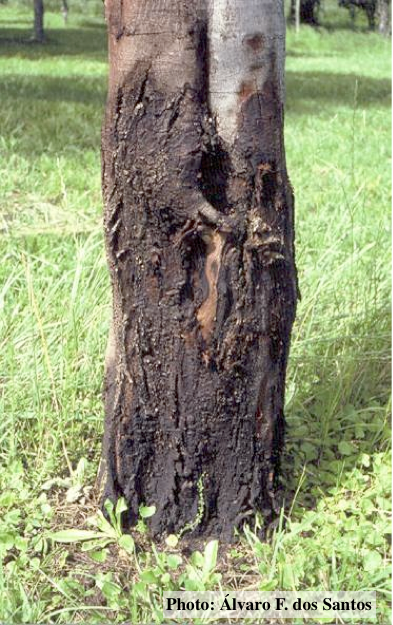 Black wattle bark with symptoms of gummosis |
|
P. cambivora sporangia 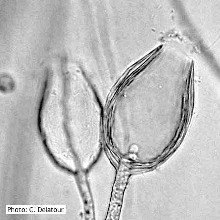 Empty sporangia of P. cambivora showing nested internal proliferation |
P. cinnamomi cork oak decline  Cork oak decline, Portugal |
P. katsurae sporangia 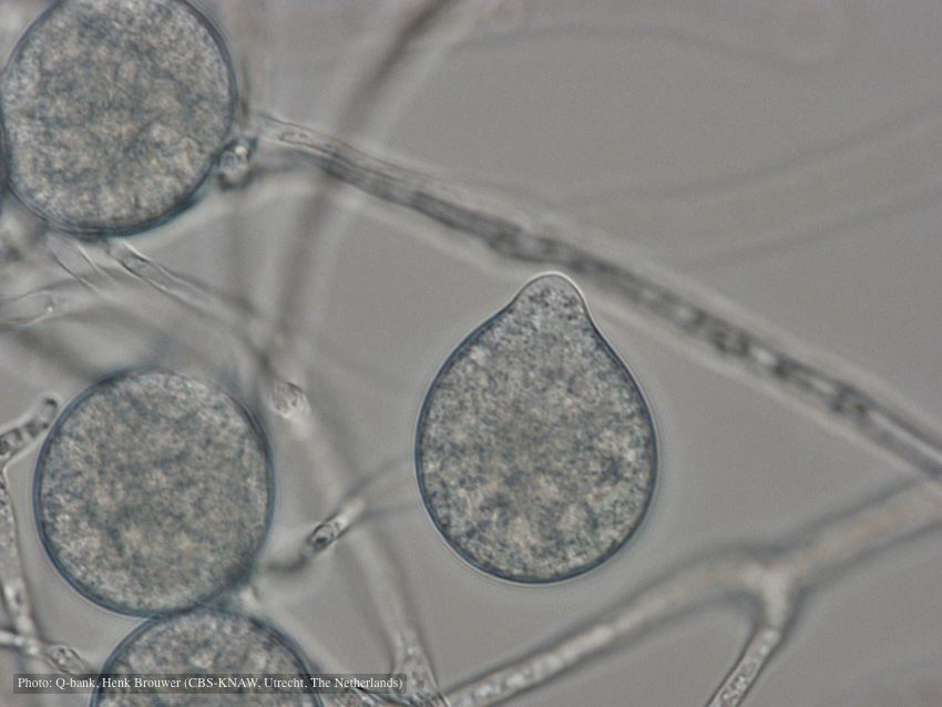 Papillate, non-caducous sporangia; photo used with permission from Q-bank |
|
P. pseudosyringae sporangia  Ovoid, semipapillate sporangia showing sympodial development of sporangiophore |
P. nemorosa sporangium  Ovoid, semi-papillate sporangium showing medium length pedicel. |
P. tentaculata disease symptoms on California mugwort  Nursery grown California mugwort plant (Artemisia douglasiana) infected with P. tentaculata and exhibiting severe root and crown rot |