

P. nicotianae overview of sporangia 40x. Photo from Q-bank: www.q-bank.eu, Henk Brouwer (CBS-KNAW, Utrecht, The Netherlands)
Photo Gallery
|
P. nicotianae sporangia  |
P. tentaculata disease symptoms on sticky monkey flower 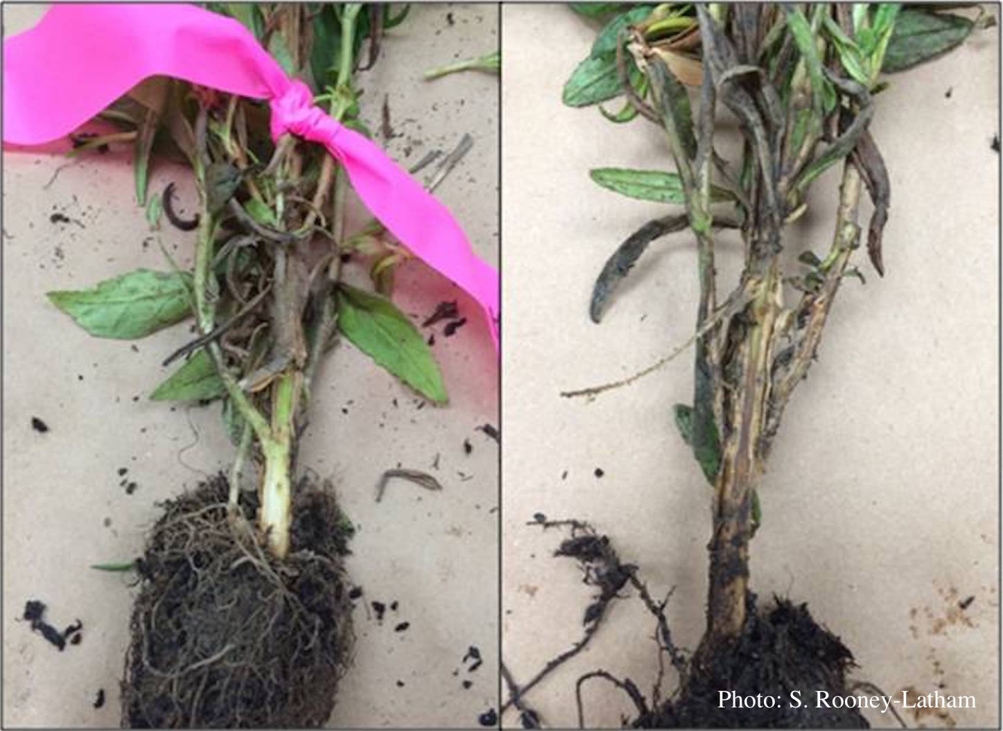 Crown and root rot (left) on sticky monkey flower (Diplacus aurantiacus) compared with a control (right) |
P. pinifolia colony morphology on CMA-NARP 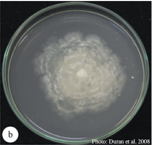 Colony morphology of P. pinifolia at 20°C on CMA-NARP after 3 weeks. From Duran et al. 2008 |
|
P. cambivora sporangia 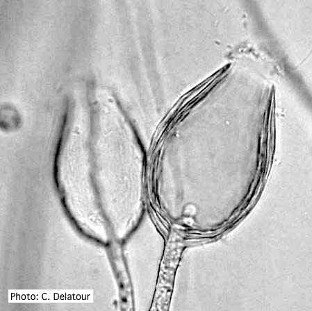 Empty sporangia of P. cambivora showing nested internal proliferation |
P. cambivora symptoms  P. cambivora on Fagus sylvatica bole |
P. alni lesion in alder, Illwald, France 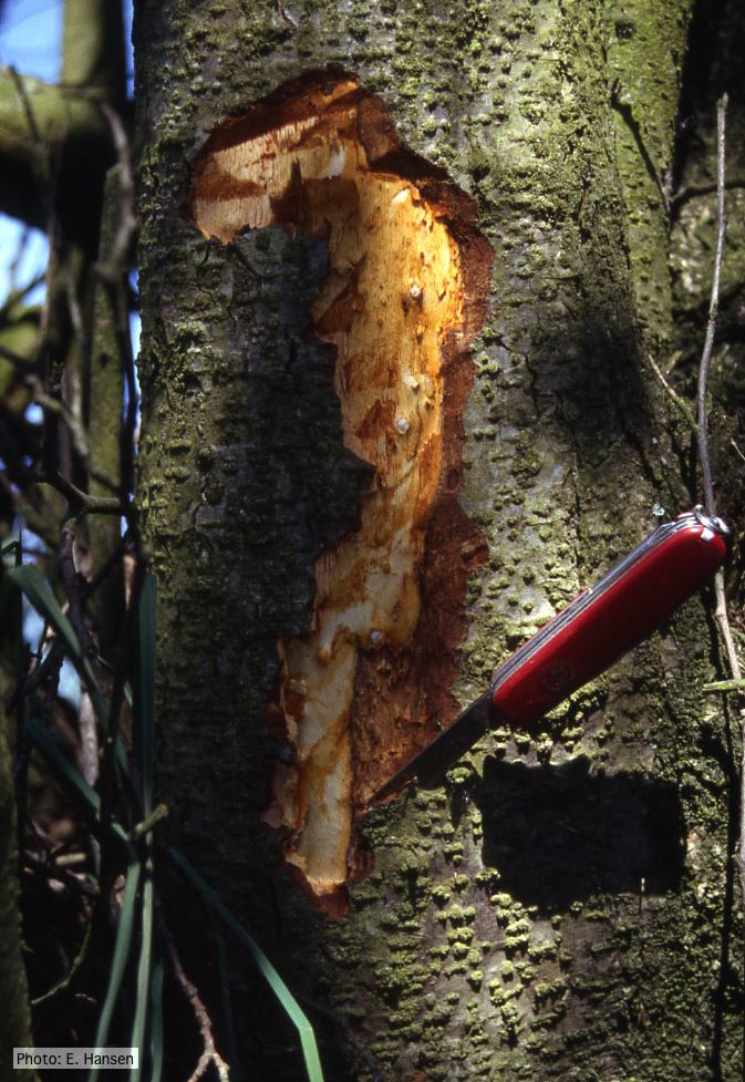 P. alni lesion in alder, Illwald, France |
|
P. pluvialis symptoms on Douglas-fir  Red needle cast symptoms on Douglas-fir in western Oregon, 2015 |
P. lateralis on Port Orford cedar  Lesion caused by aerial infection on Chaemacyparis lawsoniana in Lopérec, France |
P. pluvialis on Pinus radiata needle  Clusters of sporangia emerge from stomata of an infected radiata pine needle. |
|
P. cactorum colony morphology on V8  Colony morphology on V8 at 14 days |
P. tentaculata sporangium  Papillate sporangium of P. tentaculata |
P. katsurae oogonium  Micrograph of warty oogonium |