

Non- papillate and caducous sporangia of Phytophthora pinifolia isolated from the infected P. radiata needles.
Photo Gallery
|
P. pinifolia sporangia 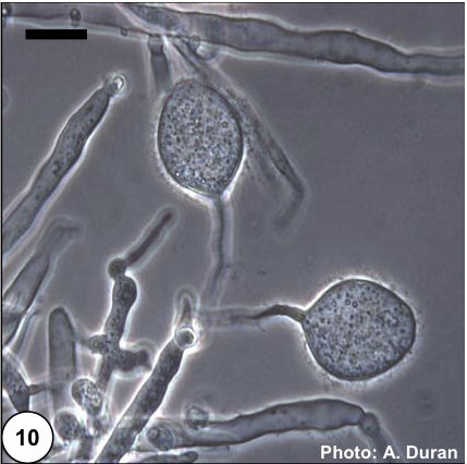 |
P. austrocedrae colony morphology on Tomato juice agar 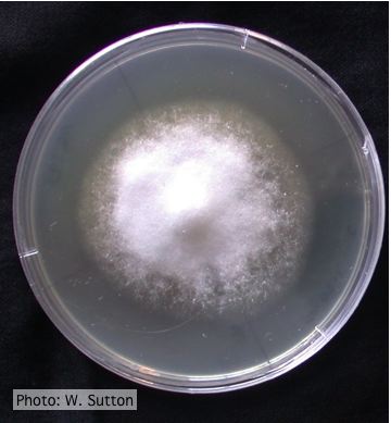 Colony morphology of P. austrocedrae at 16 ºC after 4 weeks on Tomato juice agar |
P. pluvialis on Pinus radiata in New Zealand 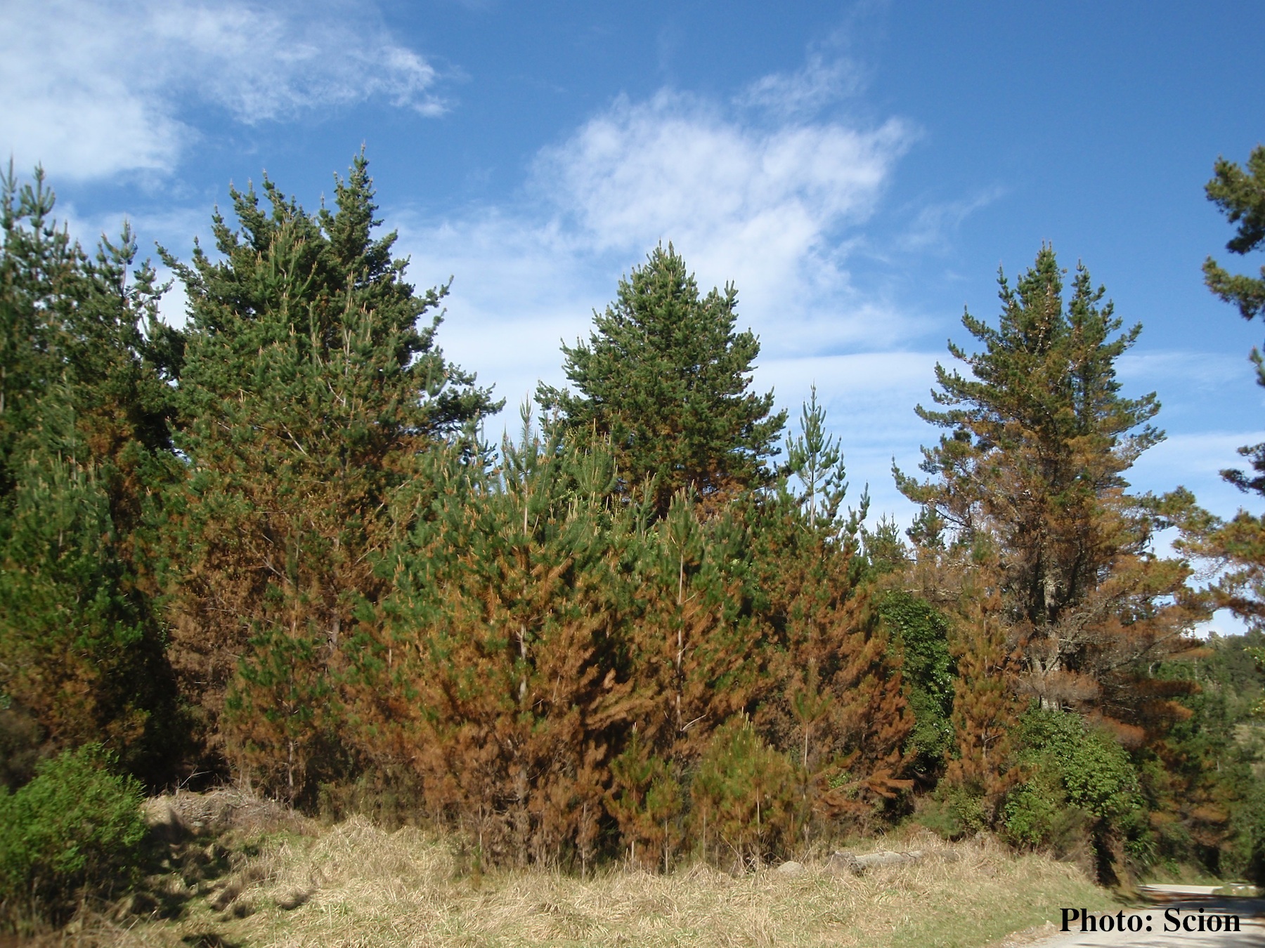 A stand of Pinus radiata trees affected by red needle cast disease. Note the reddish appearance of affected trees prior to needle drop. |
|
P. pluvialis colony morphology on carrot agar  Colony morphology on carrot agar at 20 days |
P. cinnamomi on Banksia 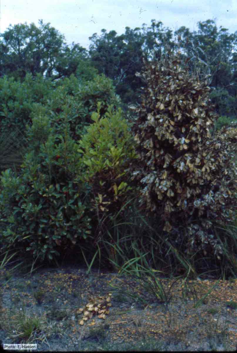 Dead and dying Banksia, Western Australia |
Comparative gametangial morphology of Phytophthora Clade 5 species  Comparative gametangial morphology of Phytophthora Clade 5 species, with SEM (top) and light microscopy (bottom). P. heveae has smooth walled oogonia with funnel-shaped, amphigynous antheridia. P. agathidicida has mildly stipulate oogonia with globose amphigynous antheridia. P.cocois has mildly bullate oogonia with reflexed amphigynous antheridia. P. castaneae has coarsely bullate oogonium with rugose protuberances and narrow amphigynous antheridia (Weir et al. 2015). |
|
P. frigida symptoms 3 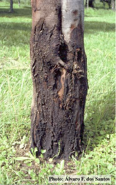 Black wattle bark with symptoms of gummosis |
P. cambivora on dead and dying chinquapin  Dead and dying chinquapin infected with P. cambivora |
P. megasperma oogonium 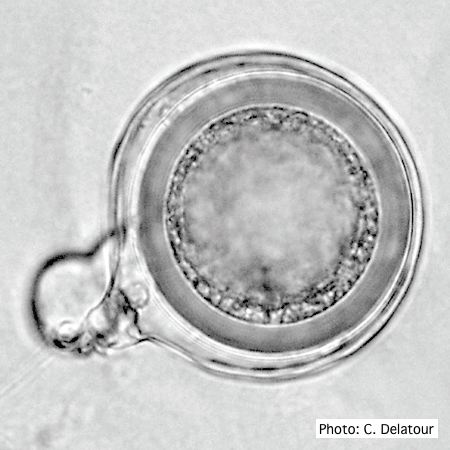 Oogonium with paragynous antheridia applied close to the ogonial stalk. |
|
P. austrocedrae colony morphology on Tomato juice agar with B sitosterol 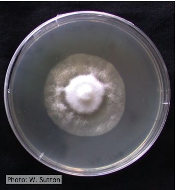 Colony morphology of P. austrocedrae at 16 ºC after 4 weeks on Tomato juice agar with B sitosterol |
P. pinifolia on Pinus radiata 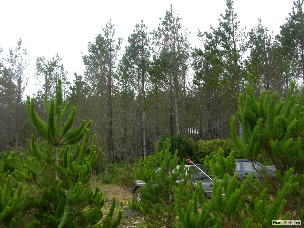 Pinus radiata stand, note Defoliation and regrowth |
P. austrocedrae - hyphal swellings  Morphology of hyphae of Phytophthora austrocedrae, from Greslebin et al. 2007 |