

Raised beds for testing disease resistance of Port-Orford-cedar seedlings at the Dorena Genetic Resource Center
Photo Gallery
Site will be retired 9/1/2026
This site is no longer being developed and will be retired on September 1, 2026. Please contact us if you have any questions or would like to provide support to continue the project.
|
Port Orford cedar seedlings 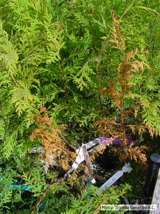 |
P. chlamydospora sporangium  Phytophthora chlamydospora sporangium in water. Bar is 20µm. |
P. arenaria disease symptoms on Banksia landscape 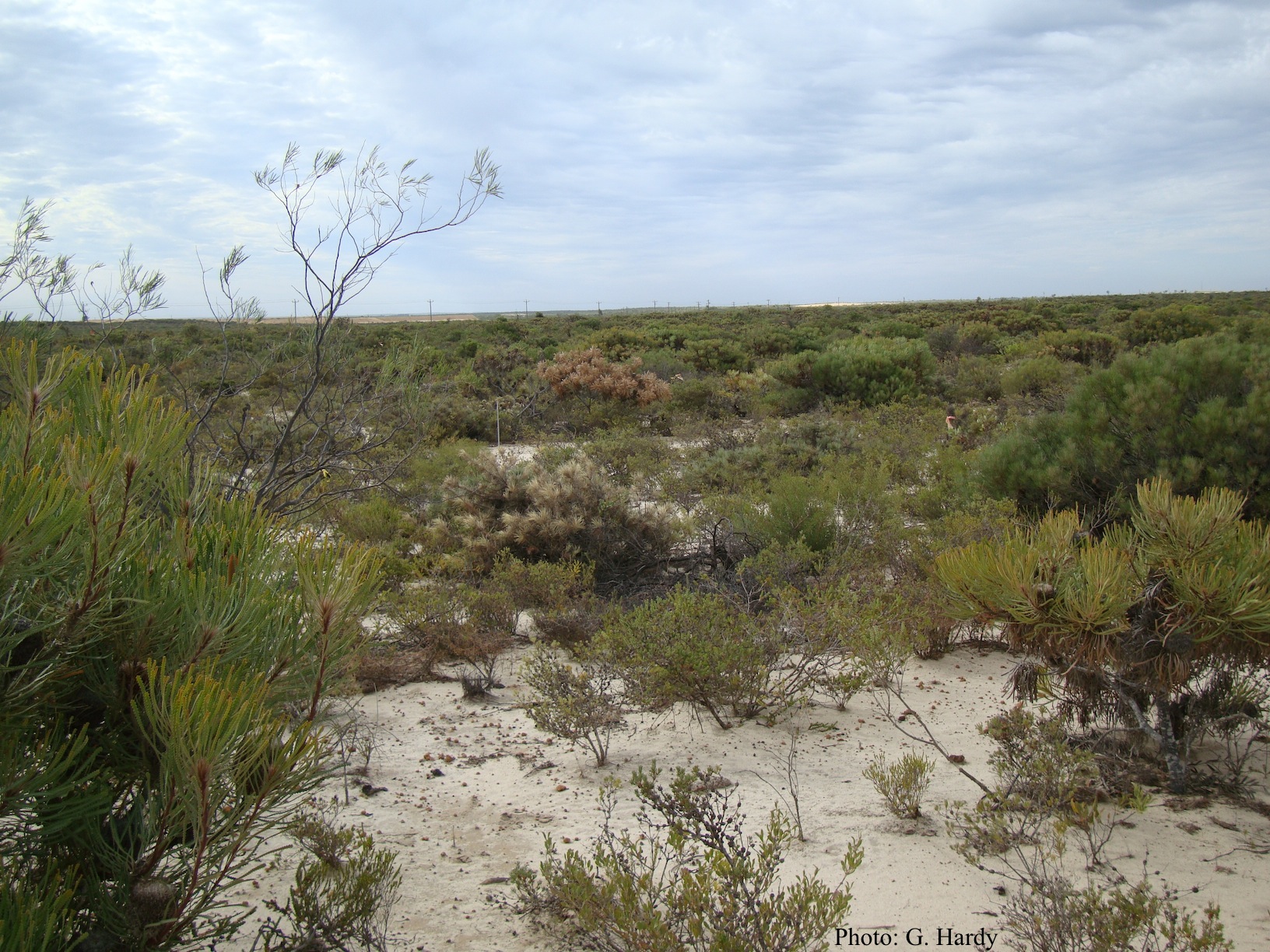 Dead Banksia sp. in a Kwongan heathland on mineral sand near Eneabba, Western Australia recently killed by root and collar rot caused by Phytophthora arenaria |
|
P. pseudotsugae colony morphology on V8 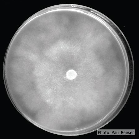 P. pseudotsugae colony growth on V8 agar |
P. frigida oogonium  Oogonium and oospore with amphigynous antheridium |
Dead Port Orford Cedar 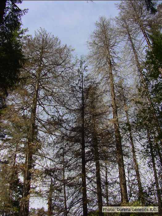 Dead Chamaecyparis lawsoniana, BLM Roseburg District in Oregon |
|
P. cryptogea colony morpholgy on V8  Colony morphology on V8 at 14 days |
P. cinnamomi on Banksia 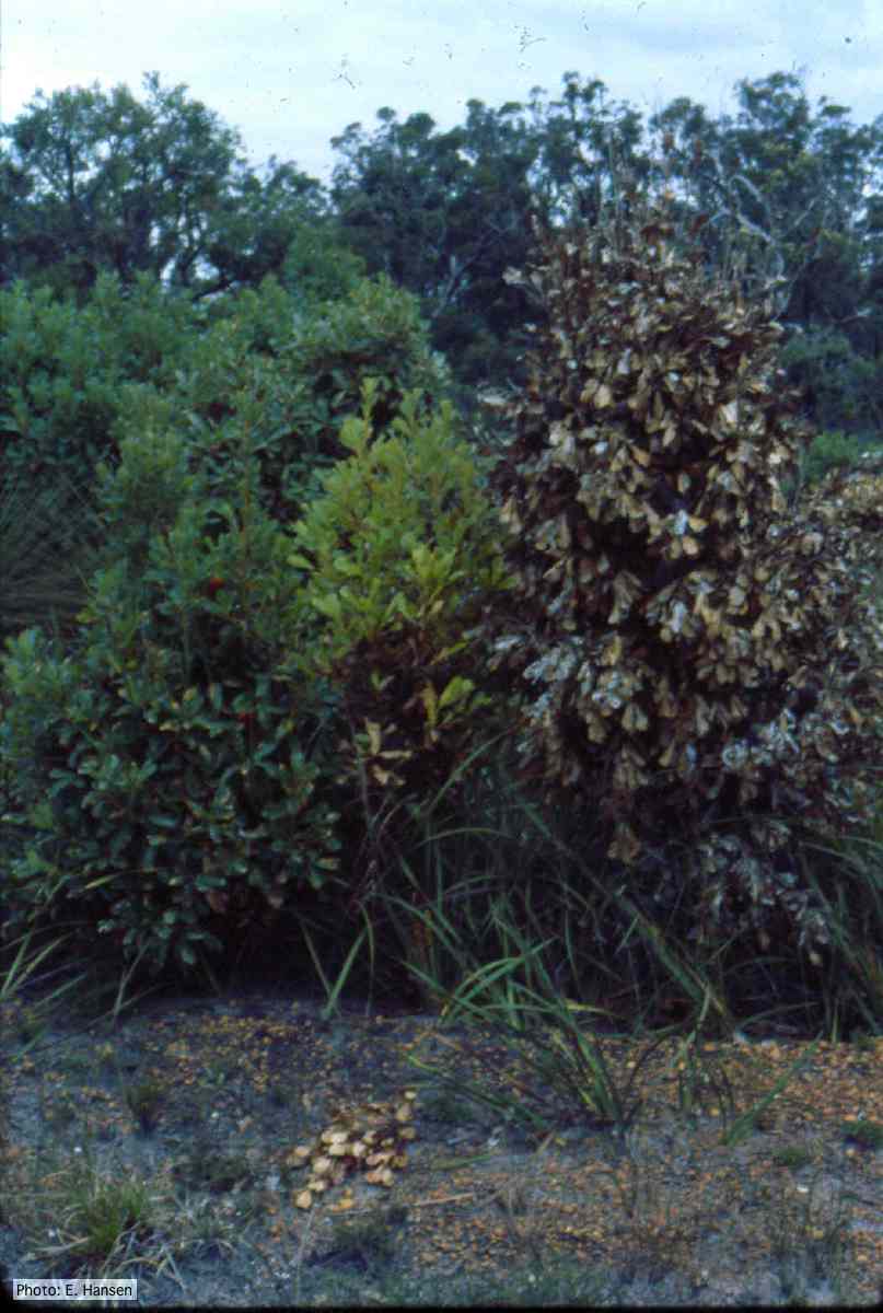 Dead and dying Banksia, Western Australia |
P. austrocedrae hyphal swellings in liquid media drawing  Morphology of hyphae of Phytophthora austrocedrae, from Greslebin et al. 2007 |
|
P. pluvialis on Pinus radiata 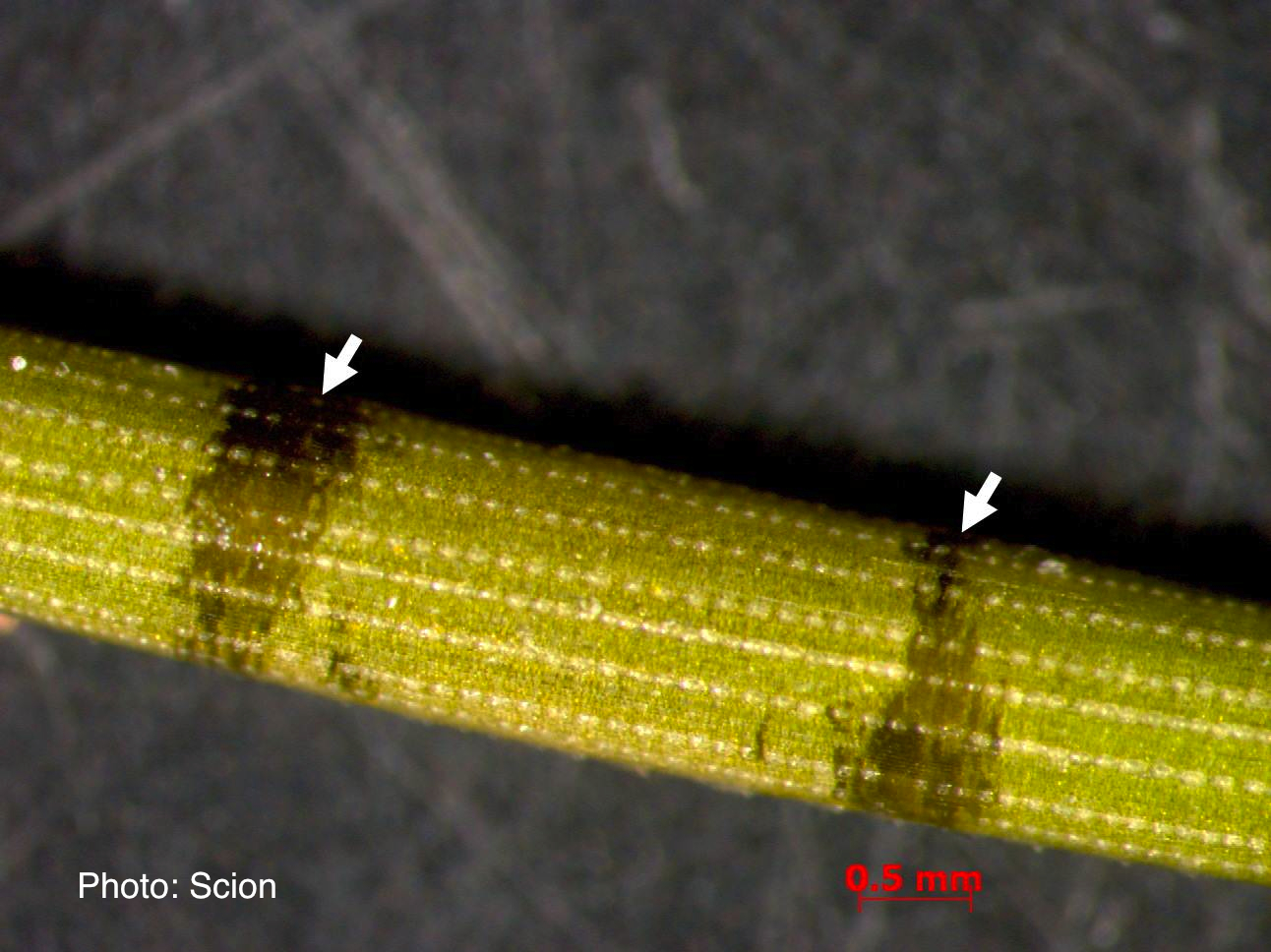 A Pinus radiata needle showing black resinous bands or marks consistent with the presence of red needle cast disease. |
P. austrocedrae - Mal del ciprés, stages of decline  Mal del ciprés, stages of decline |
P. pinifolia colony morphology on CMA-NARP 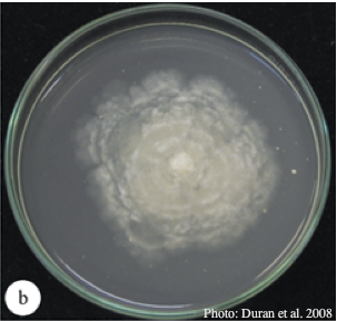 Colony morphology of P. pinifolia at 20°C on CMA-NARP after 3 weeks. From Duran et al. 2008 |