

Phytophthora collar rot on Italian alder trees: standing, dead tree
Photo Gallery
Site will be retired 9/1/2026
This site is no longer being developed and will be retired on September 1, 2026. Please contact us if you have any questions or would like to provide support to continue the project.
|
P. siskiyouensis disease symptoms on Italian alder 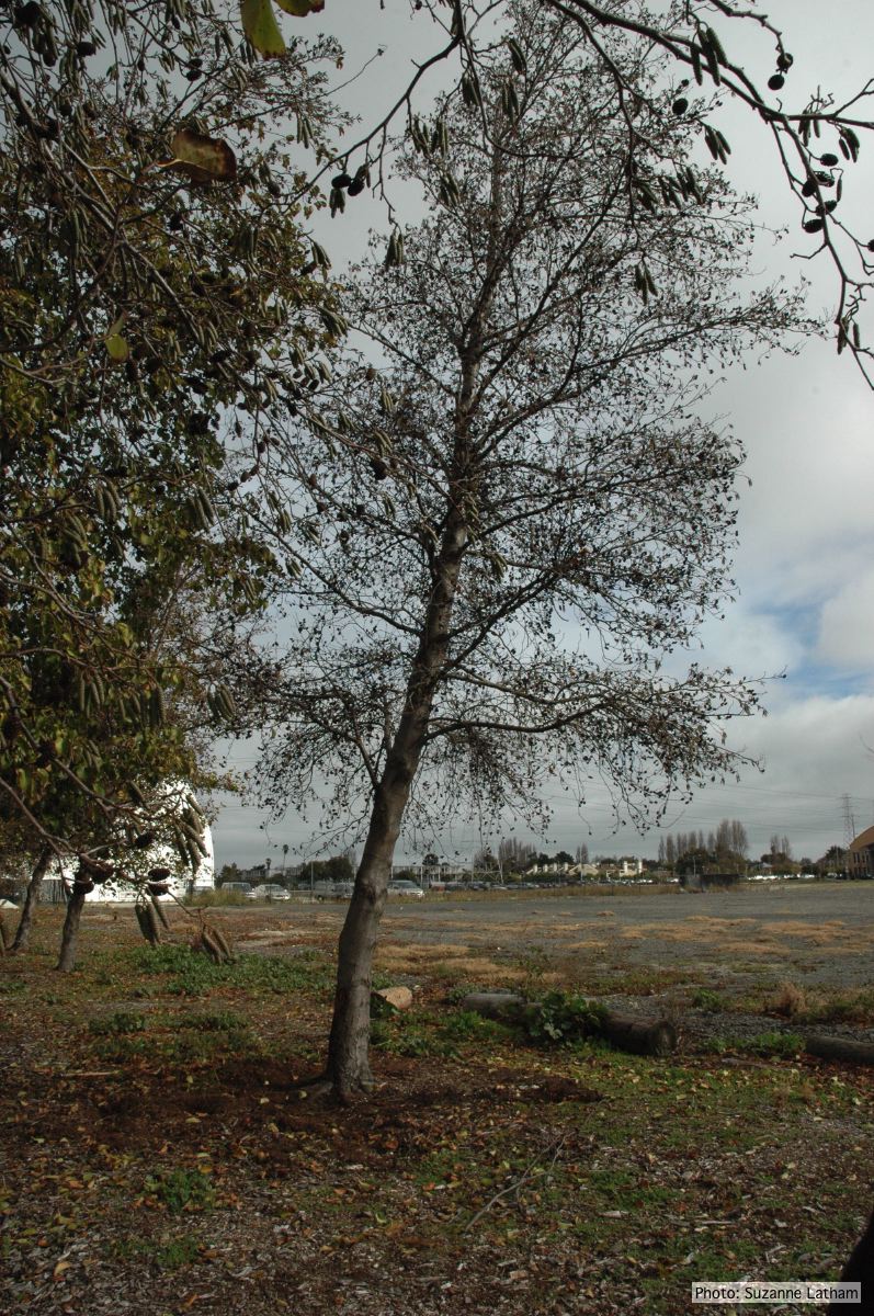 |
P. siskiyouensis bleeding canker 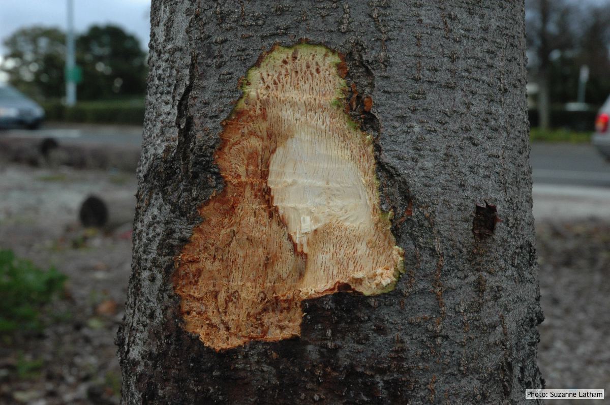 Close-up of margin area of bole lesions under the bark of a bleeding canker |
P. siskiyouensis bleeding canker  Bole lesions in the tissues under the bark of a bleeding canker: discoloration in the secondary phloem tissue |
|
P. siskiyouensis colony morphology on V8 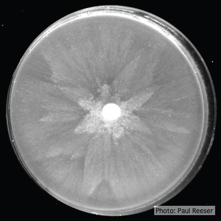 Colony morphology on V8 at 14 days |
P. siskiyouensis disease symptoms on Italian alder 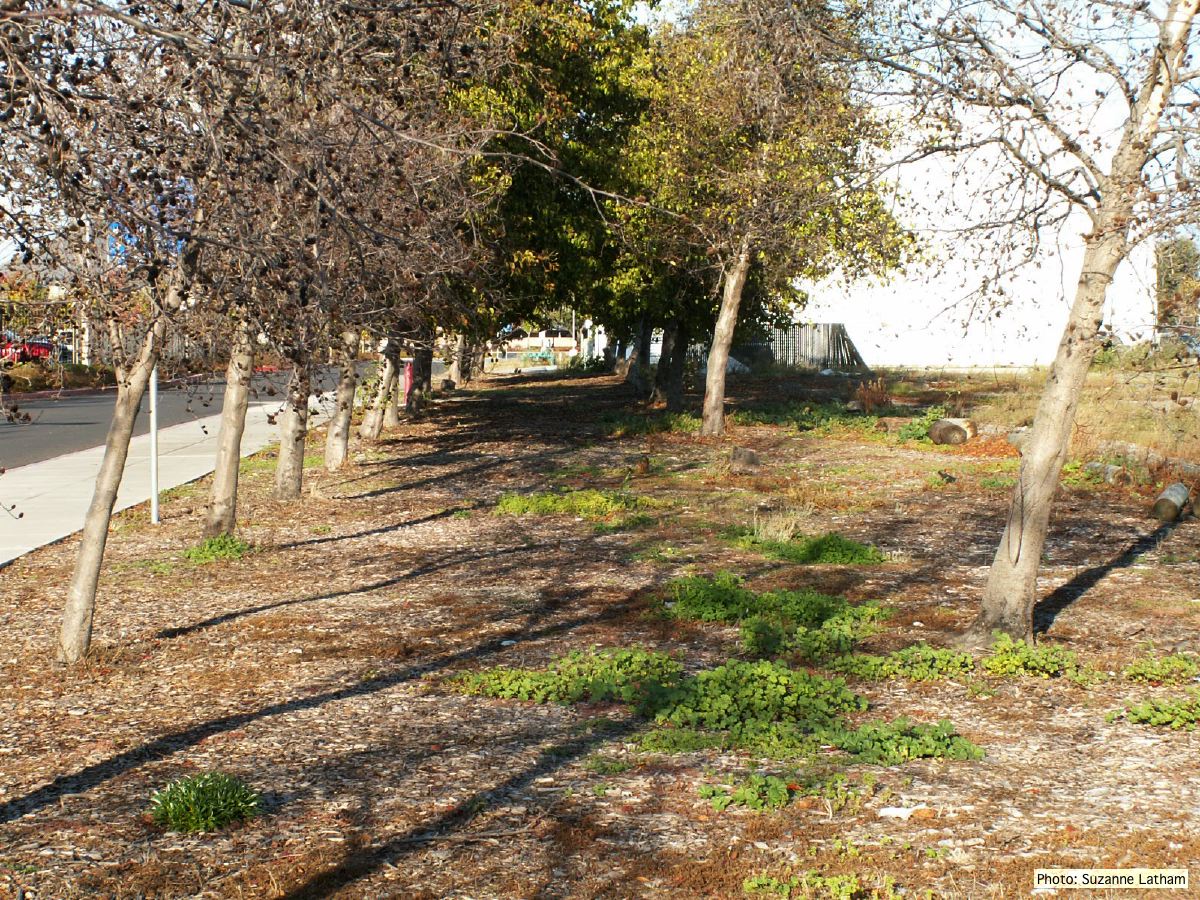 Grove of dying trees in a commercial landscape in Foster City, CA |
P. siskiyouensis sporangia 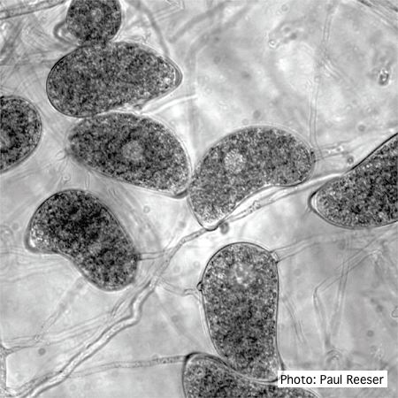 Sporangia showing a variety of shapes and orientations of semi-papillae and sporangiophores |
|
P. siskiyouensis oogonium with amphigynous antheridium 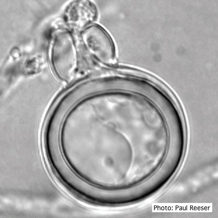 P. siskiyouensis oogonium with amphigynous antheridium |
P. siskiyouensis sporangium  P. siskiyouensis sporangium with lateral semi-papilla and subterminal, sub-basal insertion in the sporangiophore |
P. siskiyouensis oogonium with paragynous antheridium  P. siskiyouensis oogonium with paragynous antheridium |
|
P. siskiyouensis canker on Italian alder  Bleeding canker at the base of a tree and a sprinkler emitter (arrow) adjacent to the trunk |
P. siskiyouensis canker on Italian alder 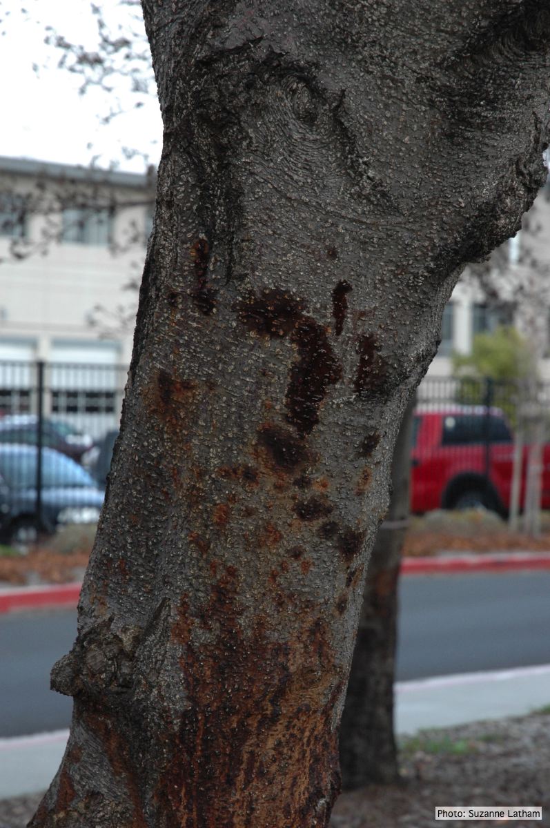 Phytophthora collar rot on Italian alder trees: an isolated bleeding canker on the trunk |
P. siskiyouensis colony morphology on PDA 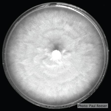 Colony morphology on PDA at 14 days |