

Non- papillate and caducous sporangia of Phytophthora pinifolia isolated from the infected P. radiata needles.
Photo Gallery
|
P. pinifolia sporangium 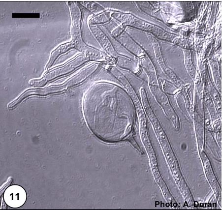 |
P. pinifolia on Pinus radiata 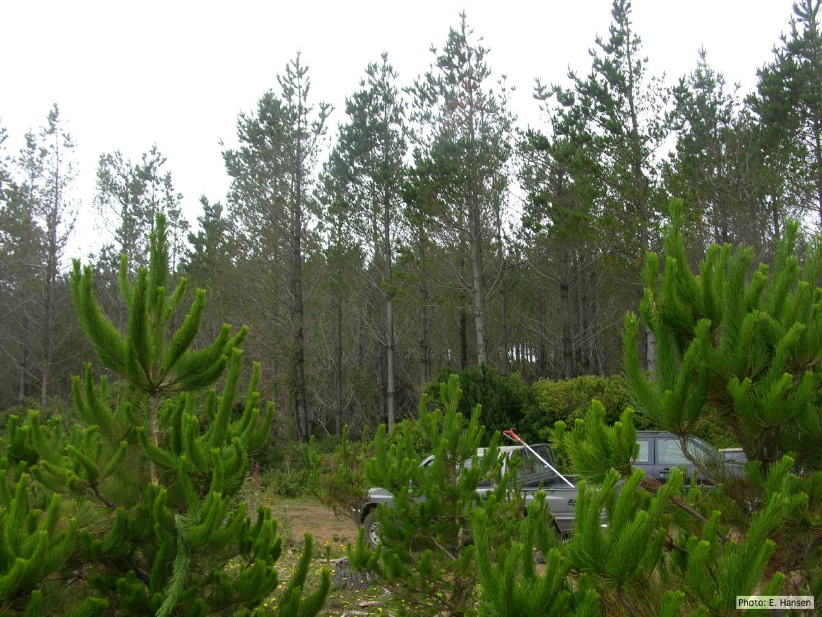 Pinus radiata stand, note Defoliation and regrowth |
P. pinifolia colony morphology on CMA-NARP 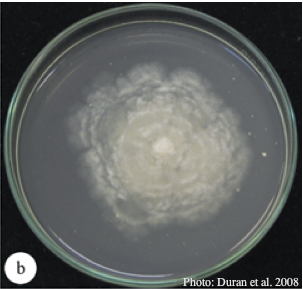 Colony morphology of P. pinifolia at 20°C on CMA-NARP after 3 weeks. From Duran et al. 2008 |
|
P. pinifolia hyphal growth 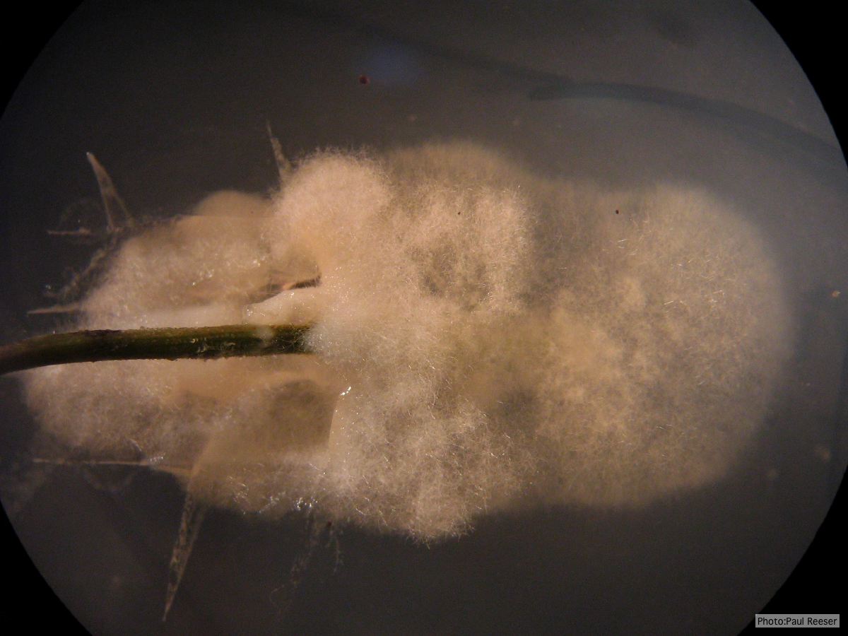 P. pinifolia pathogen growing from infected needle on selective agar |
P. pinifolia on Pinus radiata 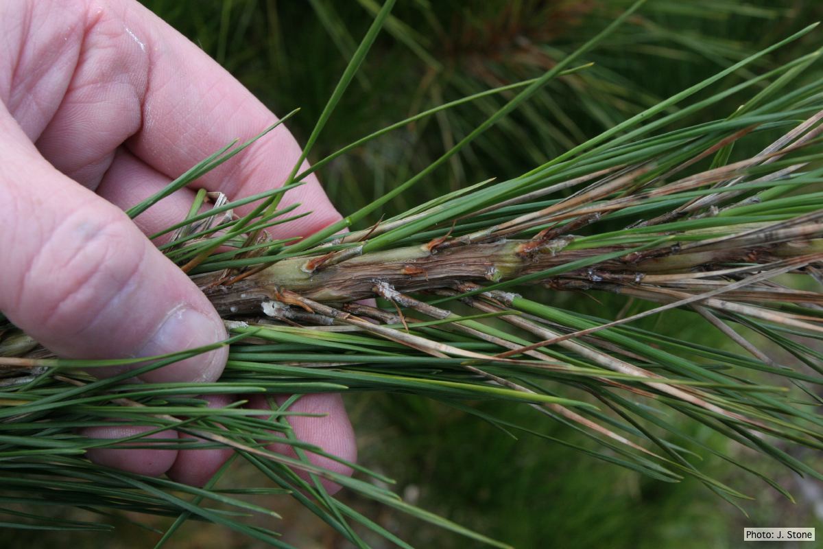 Pinus radiata needles, note “black line” symptom near needle bases |
P. pinifolia sporangia 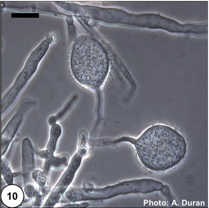 Non- papillate and caducous sporangia of Phytophthora pinifolia isolated from the infected P. radiata needles. |
|
P. pinifolia sporangia 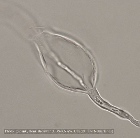 Sporangium with internal proliferation, photo from Q-bank, used with permission. |
P. pinifolia colony morphology on V8 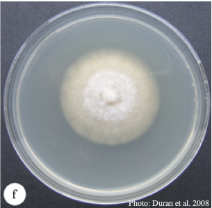 Colony morphology of P. pinifolia at 20°C on V8 after 3 weeks. From Duran et al. 2008 |
P. pinifolia on Pinus radiata  Pinus radiata, note Stem canker associated with necrotic needles. |
|
P. pinifolia on Pinus radiata 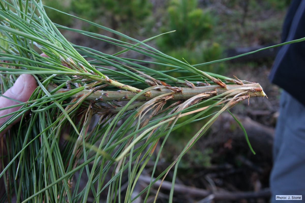 Pinus radiata, note grey and collapsed needle bases |