

Phytophthora chlamydospora chlamydospore in agar. Bar is 20µm.
Photo Gallery
|
P. chlamydospora chlamydospore  |
P. austrocedrae semipapillate sporangium  P. austrocedrae - semipapillate sporangium with off-center attachment. |
P. austrocedrae colony morphology on CMA 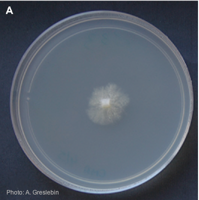 Colony morphology of P. austrocedrae at 16ºC after 4 weeks on CMA |
|
P. chlamydospora sporangium  Phytophthora chlamydospora sporangium in water. Bar is 20µm. |
P. alni sporangia 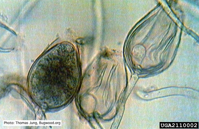 Non-papillate sporangia of P. alni showing nested proliferation. |
P. agathidicida sporangium 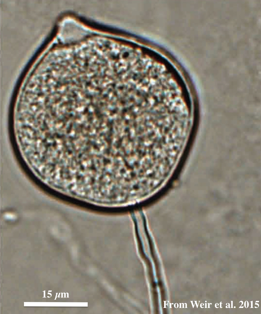 Globose to ovoid-ellipsoid, papillate sporangium |
|
P. pseudotsugae colony morphology on V8 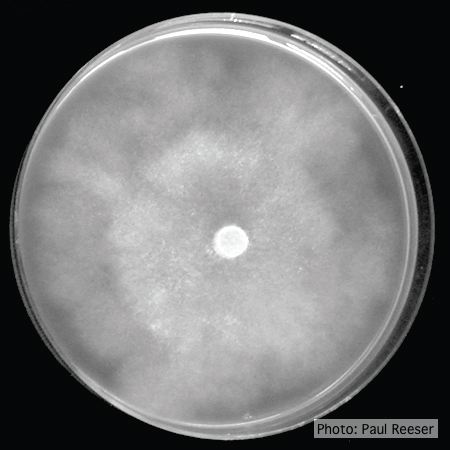 P. pseudotsugae colony growth on V8 agar |
Growth morphology on V8 of P. lateralis 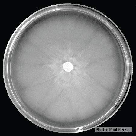 Colony morphology on V8 at 14 days |
P. cinnamomi cork oak decline  Cork oak decline, Portugal |
|
P. siskiyouensis colony morphology on PDA 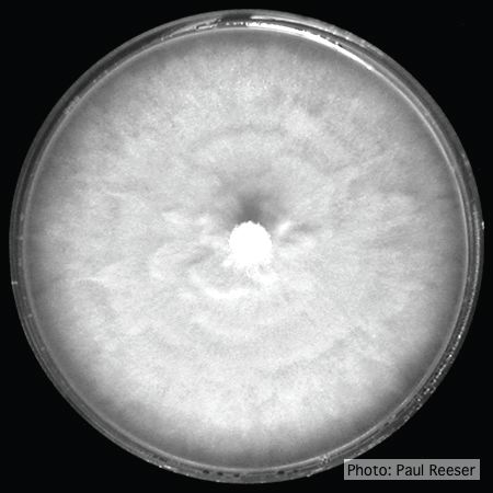 Colony morphology on PDA at 14 days |
P. tentaculata disease symptoms on California mugwort 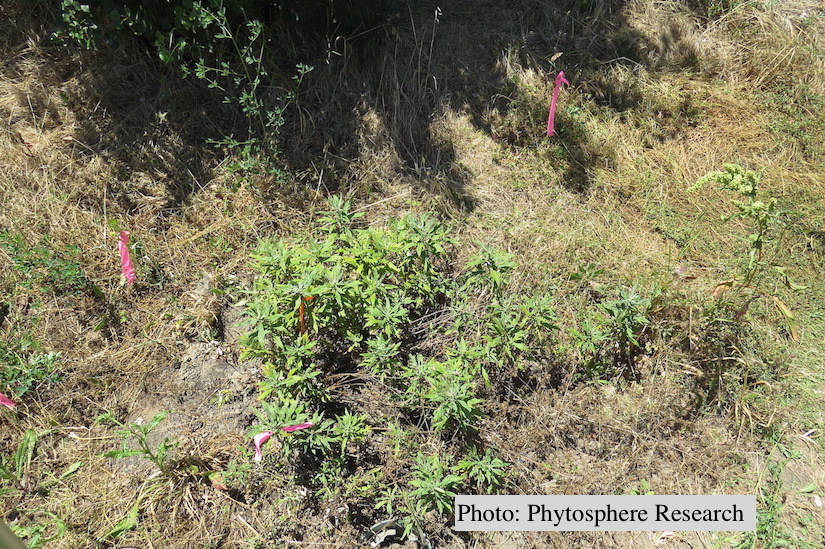 Outplanted California mugwort (Artemisia douglasiana) infected with P. tentaculata, 4.5 years after planting. Plant shows stunting and chlorosis. (P. cryptogea and P. lacustris were also baited from roots/soil of this plant). |
P. cambivora on dead and dying chinquapin  Dead and dying chinquapin infected with P. cambivora |