

P. cinnamomi hyphal swellings (or thin walled chlamydospores)
Photo Gallery
|
P. cinnamomi hyphal swellings 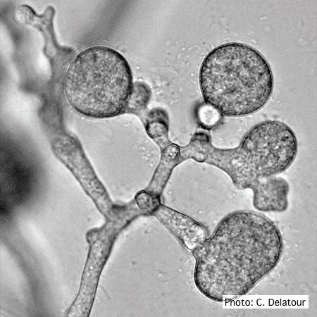 |
P. pluvialis sporangia.  P. pluvialis sporangia on tape peel from infected Douglas-fir needle. |
P. arenaria sporangia  Globose papillate sporangia of Phytophthora arenaria on V8 agar flooded with soil extract. (Scale bar = 20 μm) |
|
P. chlamydospora chlamydospore  Phytophthora chlamydospora chlamydospore in agar. Bar is 20µm. |
P. nicotianae symptoms 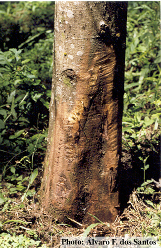 Symptoms of gummosis on black wattle (Fitopatol. bras. 2005) |
P. austrocedrae - necrotic lesion in phloem 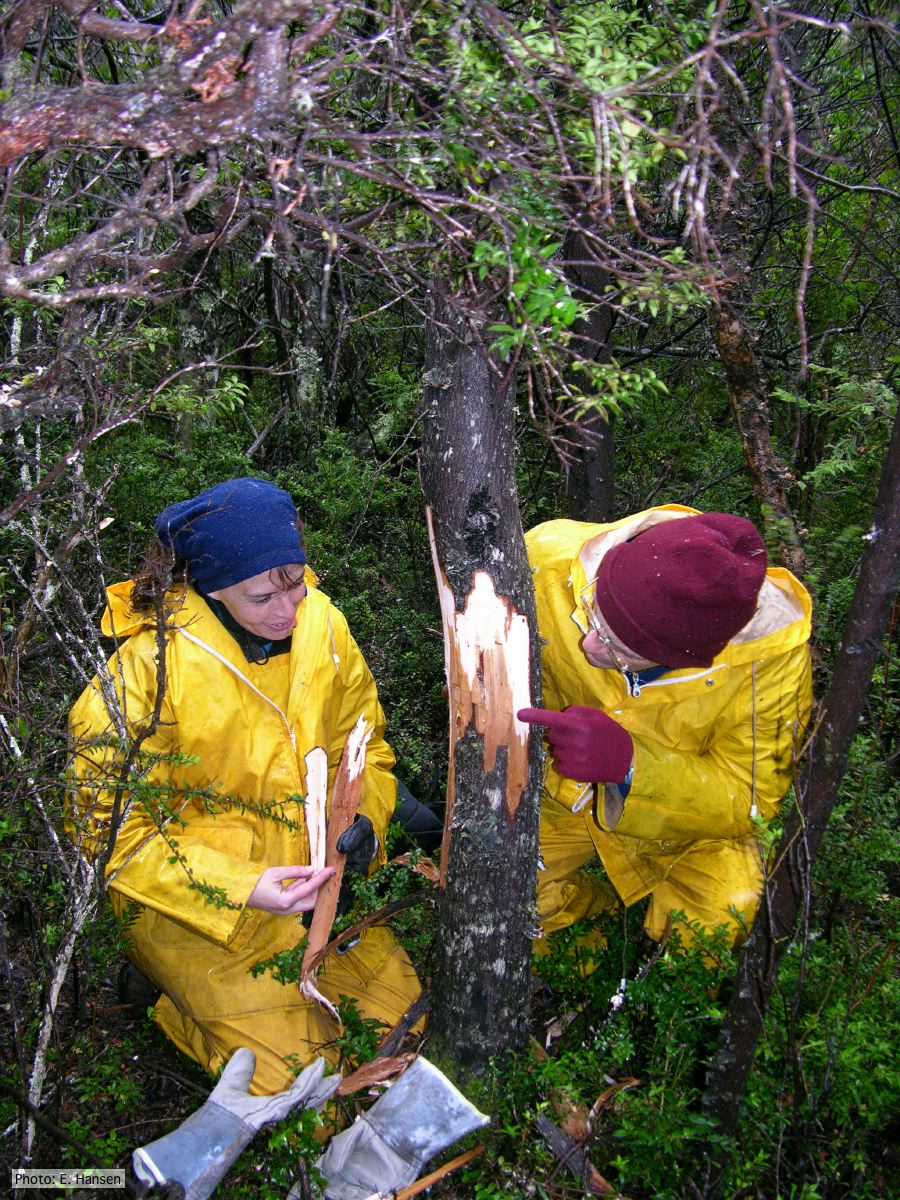 |
|
P. austrocedrae oogonia drawing  P. austrocedrae. Morphology of oogonia, oospores and antheridia. Bar: 10 mm. Greslebin et al. 2007 |
P. austrocedrae - sporangia drawings 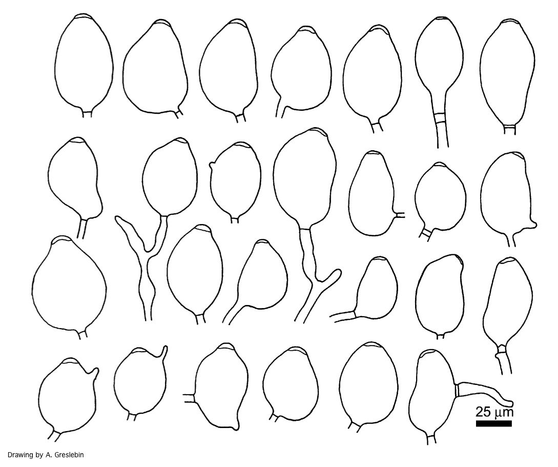 Phytophthora austrocedrae. Morphology of sporangia. Bar: 25 mm. Greslebin et al. 2007 |
P. cambivora colony morphology on MA  Appressed colony morphology at 14 days at 20°C on MA |
|
P. cambivora tar spots 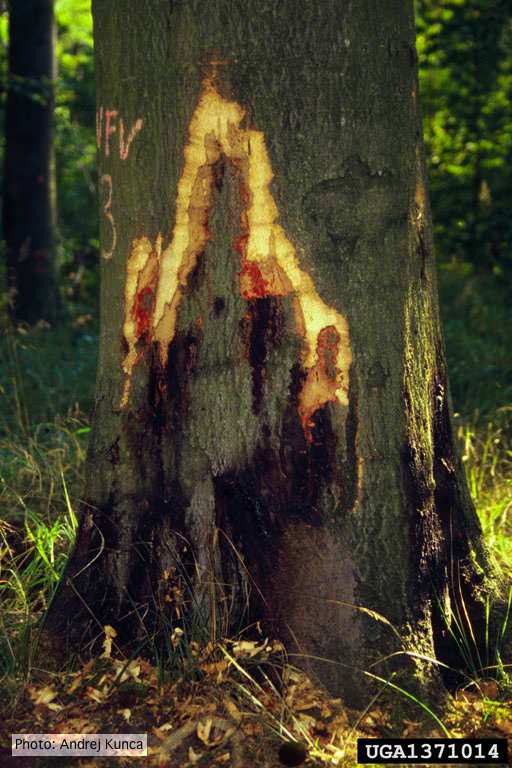 Tar spots on European beech (Fagus sylvatica) with bark removed. Lesse, Germany |
P. lateralis sporangia  Sympodial sporangiophore with external proliferation |
P. pluvialis symptoms on Douglas-fir  Red needle cast symptoms on Douglas-fir in western Oregon, 2015 |