

Colony morphology of P. pinifolia at 20°C on V8 after 3 weeks. From Duran et al. 2008
Photo Gallery
|
P. pinifolia colony morphology on V8 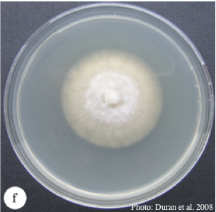 |
P. tentaculata sporangium 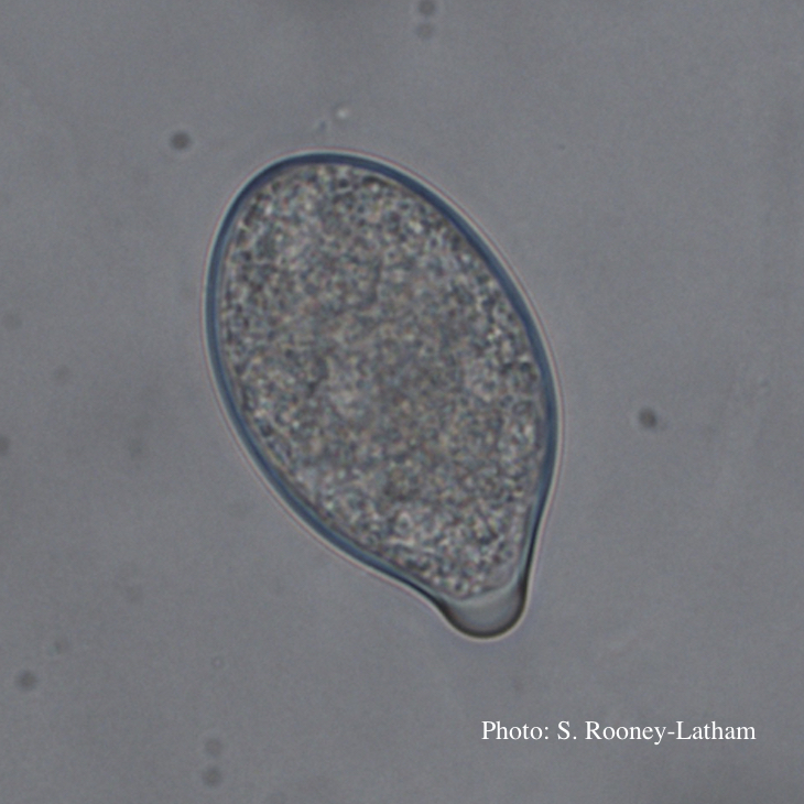 Papillate sporangium of P. tentaculata |
P. siskiyouensis colony morphology on PDA 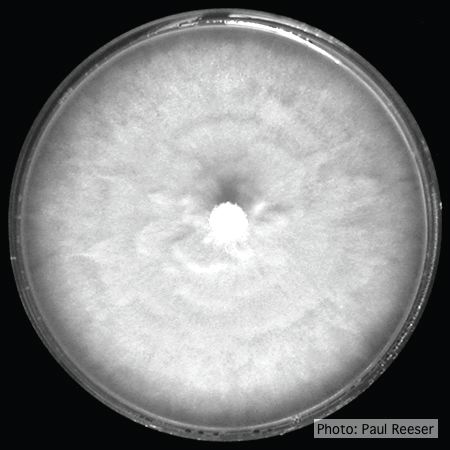 Colony morphology on PDA at 14 days |
|
P. cryptogea sporangia  Sporangiophore showing internal proliferation through empty sporangia after zoospore release |
P. agathidicia oogonia 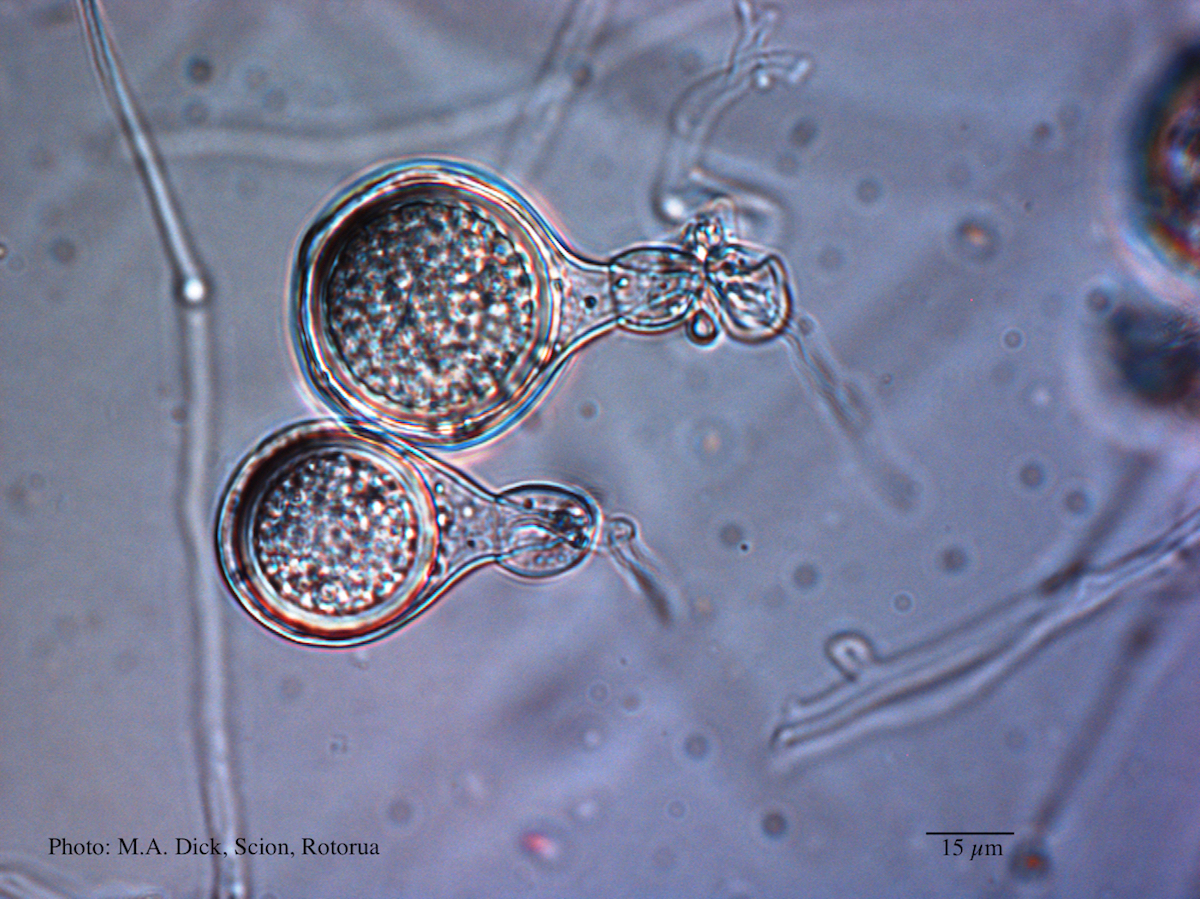 Light micrograph of P. agathidicida oospore (Scale bar equals 15 µm) |
P. pluvialis on Pinus radiata in New Zealand  Typical red needle cast symptoms along a twig. Lesions begin at the base of the needle which subsequently turns brown and is cast from the twig. |
|
P. nicotianae chlamydospore 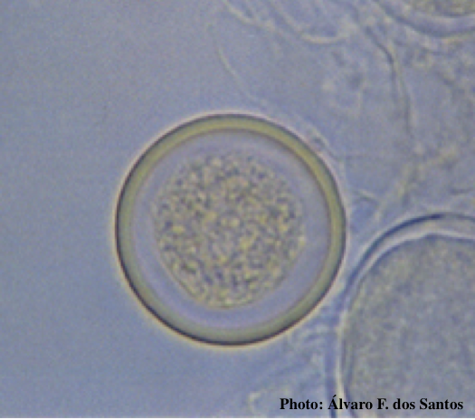 Globose chlamydospore (Fitopatol. bras. 2005) |
P. arenaria disease symptoms on Banksia landscape 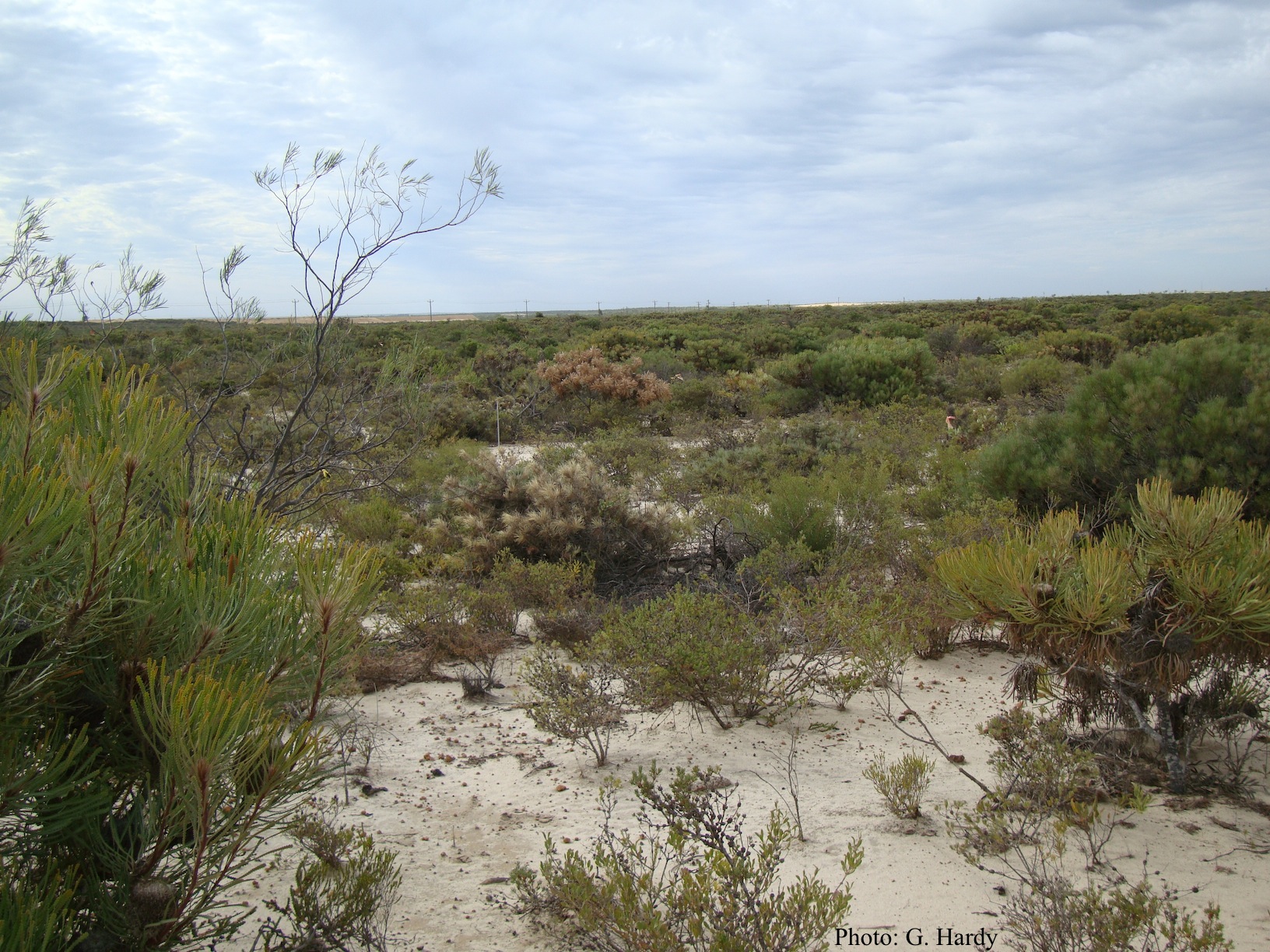 Dead Banksia sp. in a Kwongan heathland on mineral sand near Eneabba, Western Australia recently killed by root and collar rot caused by Phytophthora arenaria |
P. cactorum bleeding canker  Bleeding canker on European beech (Fagus sylvatica) |
|
P. chlamydospora colony morphology on carrot agar 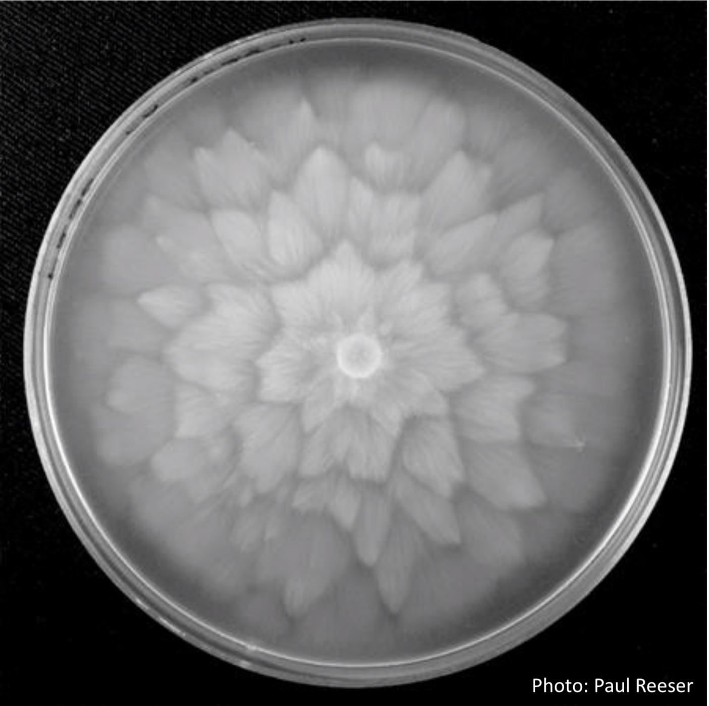 P. chlamydospora colony morphology on carrot agar |
P. tentaculata disease symptoms on sticky monkey flower 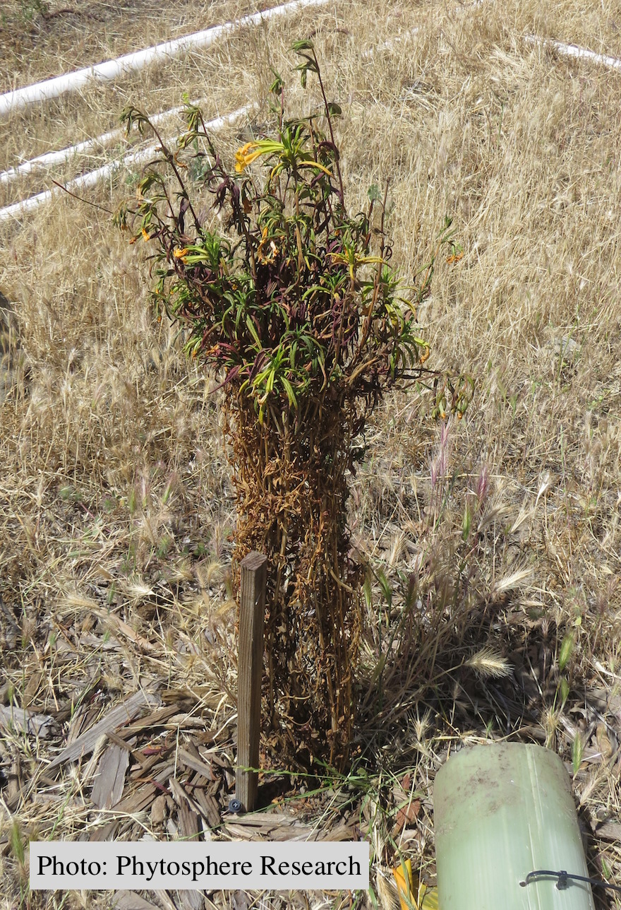 Outplanted sticky monkey flower (Diplacus aurantiacus) infected with P. |
P. cambivora coralloid irregular hyphae  Coralloid irregular hyphae |