

Phytophthora austrocedrae. Morphology of sporangia. Bar: 25 mm. Greslebin et al. 2007
Photo Gallery
|
P. austrocedrae - sporangia drawings 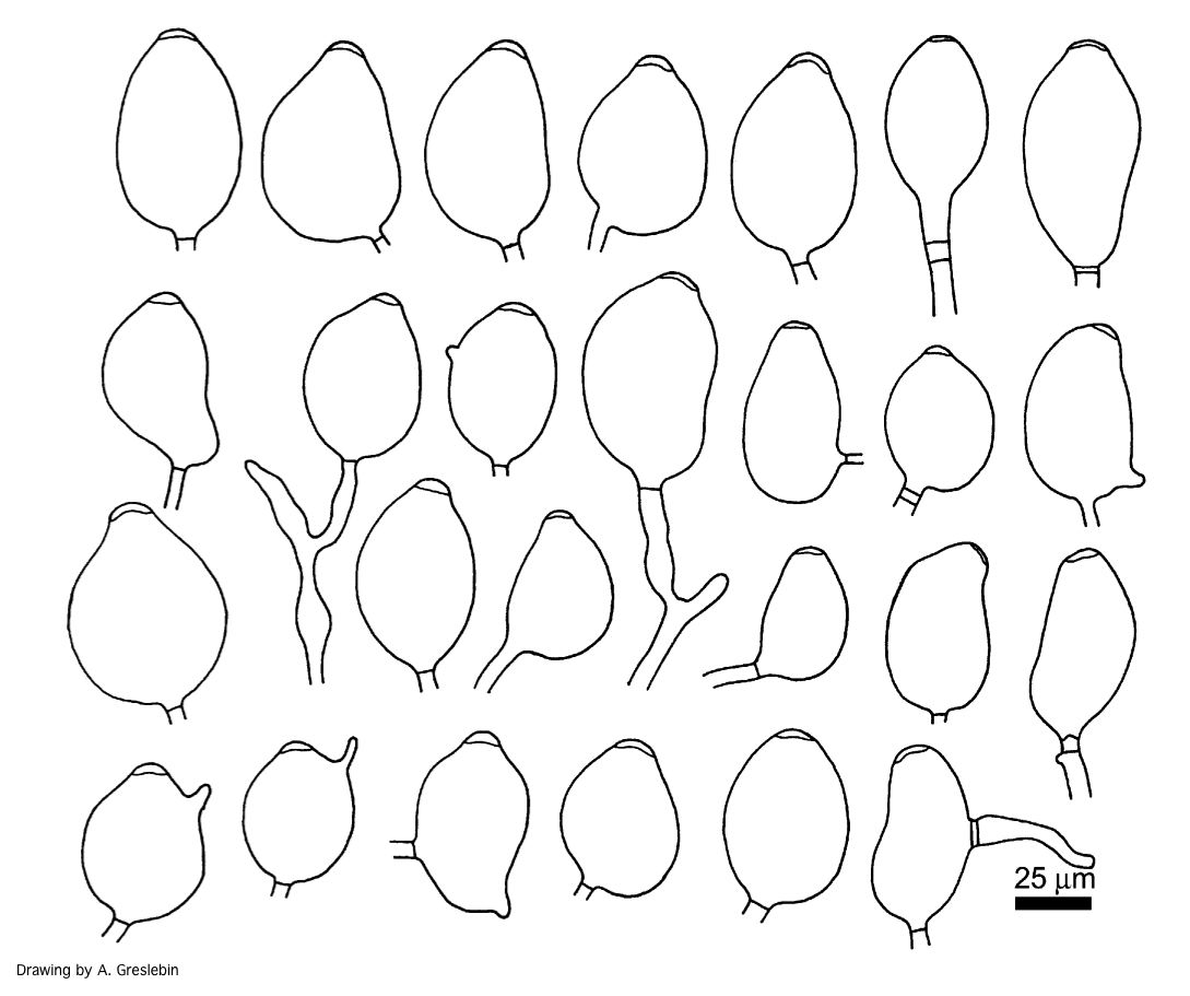 |
P. cinnamomi sporangium 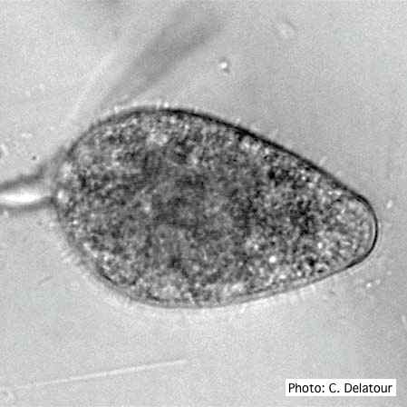 P. cinnamomi sporangium |
P. pseudotsugae colony morphology on PDA  P. pseudotsugae colony growth on PDA agar |
|
P. frigida symptoms 3 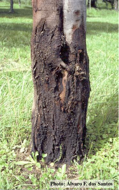 Black wattle bark with symptoms of gummosis |
P. cryptogea colony morpholgy on V8  Colony morphology on V8 at 14 days |
P. katsurae oogonium 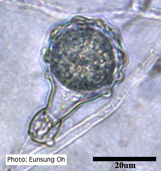 Warty protuberances on oogonium |
|
P. cambivora colony morphology on MA 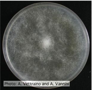 Uniform fluffy colony morphology at 14 days at 20°C on MA |
P. boehmeriae symptoms 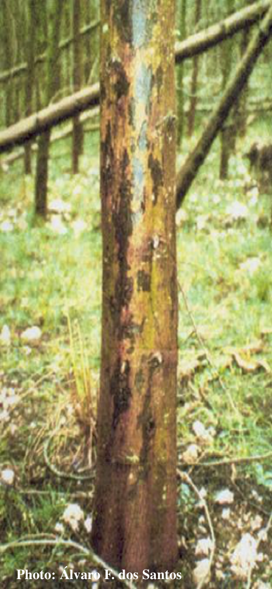 Symptoms of gummosis on black wattle (Acacia mearnsii) |
P. nemorosa sporangia  Ovoid, semi-papillate sporangia showing sympodial development of sporangiophore |
|
P. tentaculata disease symptoms on California mugwort 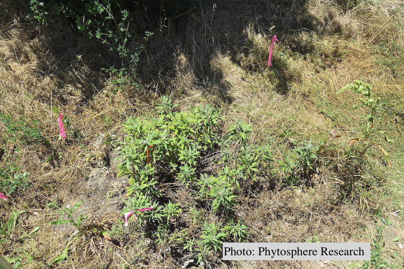 Outplanted California mugwort (Artemisia douglasiana) infected with P. tentaculata, 4.5 years after planting. Plant shows stunting and chlorosis. (P. cryptogea and P. lacustris were also baited from roots/soil of this plant). |
P. pinifolia on Pinus radiata 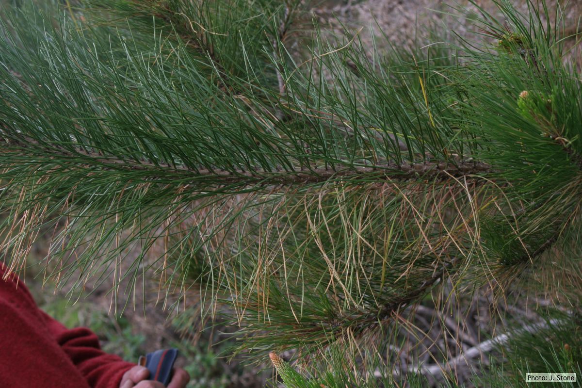 Dead needles on lower side of P. radiata branch. |
P. alni subsp alni sporangium 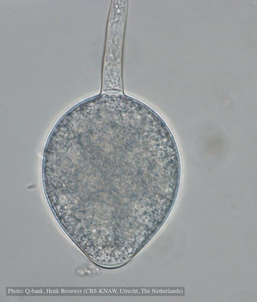 Non-papillate, non caducous sporangium, photo used with permission from Q-bank |