

Uniform fluffy colony morphology at 14 days at 20°C on PDA
Photo Gallery
|
P. cambivora colony morphology on PDA 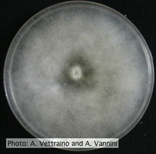 |
P. cambivora colony morphology on PDA  Cottony colony morphology at 14 days at 20°C on PDA |
P. arenaria sporangia  Globose papillate sporangia of Phytophthora arenaria on V8 agar flooded with soil extract. (Scale bar = 20 μm) |
|
P. pinifolia sporangium 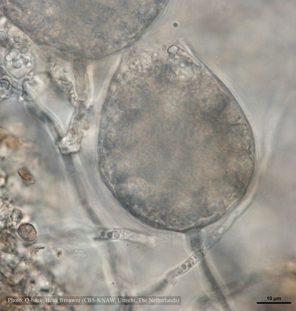 Cysts remain in sporangium after discharge, photo from Q-bank, used with permission |
P. alni symptoms on European Alder  Mature, riparian common alder (A. glutinosa) stand heavily impacted by root and collar rot caused by P. alni |
Growth of P. megakarya on PDA 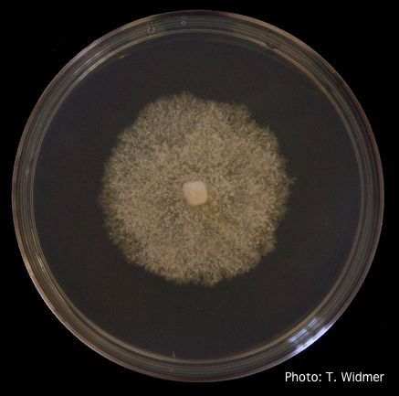 Growth of P. megakarya on potato dextrose agar |
|
P. austrocedrae - necrotic lesion in phloem 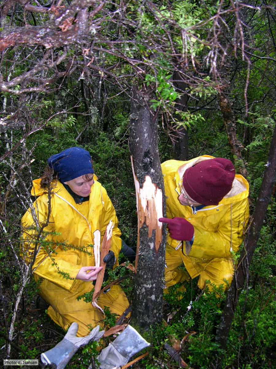 |
P. cambivora disease symptoms 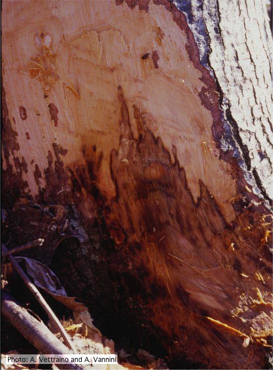 Collar canker rot of Ink disease on sweet chestnut |
P. chlamydospora hyphal swellings 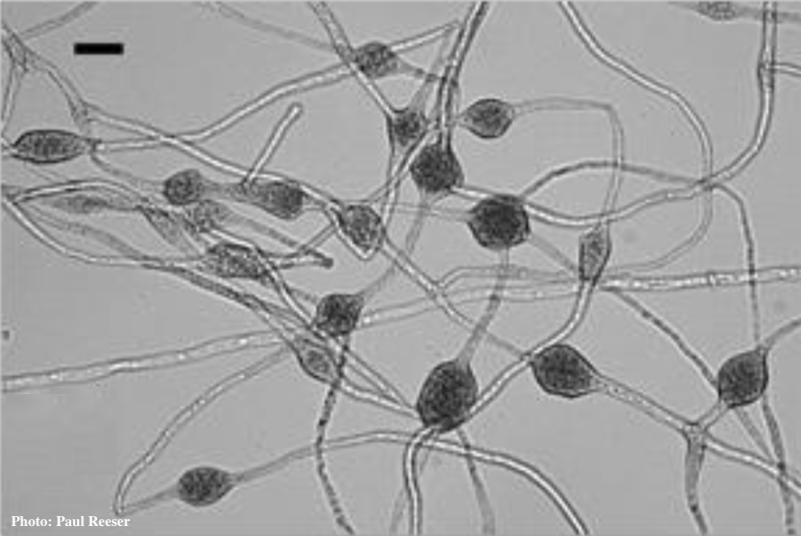 Phytophthora chlamydospora chlamydospore in agar. Bar is 20µm.
|
|
P. cinnamomi on Banksia 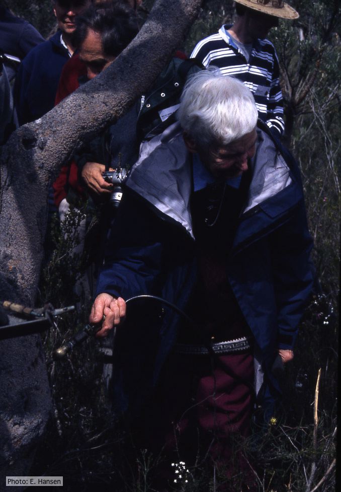 Gretna Weste injecting Banksia with phosphonate |
P. cambivora colony morphology on V8 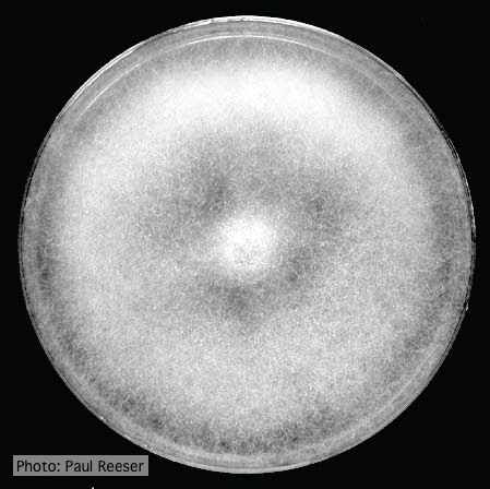 Colony morphology on V8 at 14 days |
P. cambivora tar spots 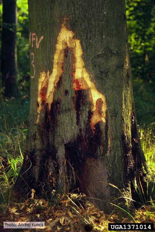 Tar spots on European beech (Fagus sylvatica) with bark removed. Lesse, Germany |