

Coralloid irregular hyphae
Photo Gallery
|
P. cambivora coralloid irregular hyphae  |
P. cambivora disease symptoms 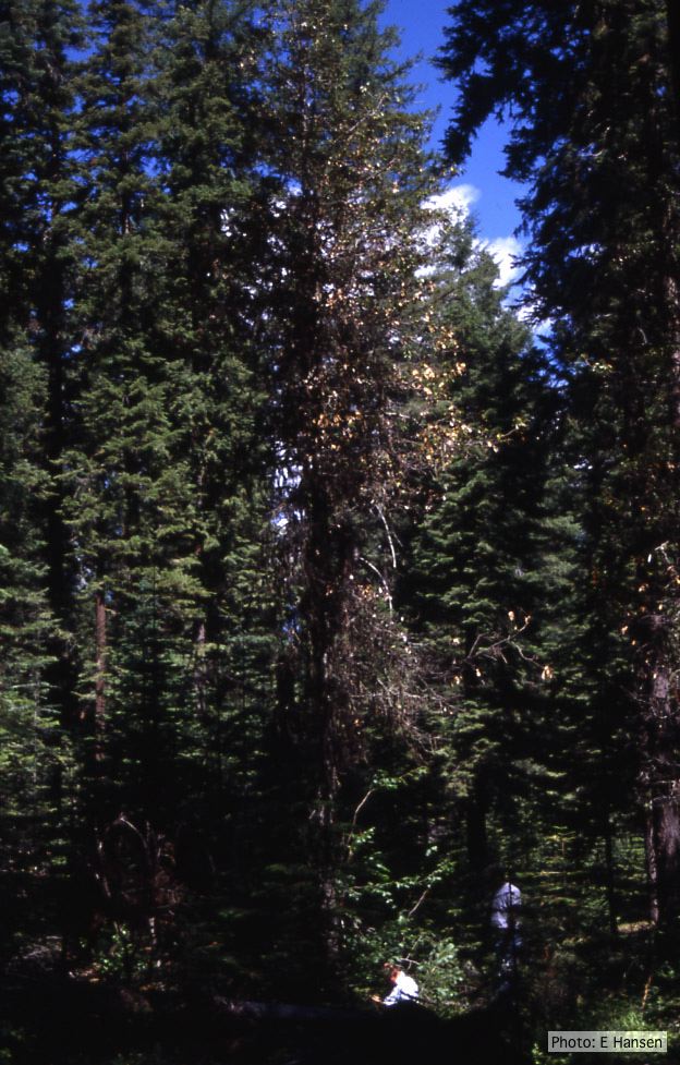 Dead and dying chinquapin infected with P. cambivora |
P. cambivora sporangium 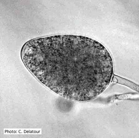 Ovoid non- papillate sporangia |
|
P. cambivora colony morphology on MA 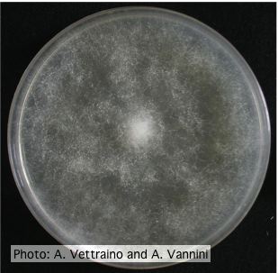 Uniform fluffy colony morphology at 14 days at 20°C on MA |
P. cambivora oogonium 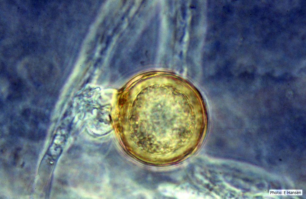 P. cambivora oogonium with antheridium |
P. cambivora inactive lesion on chinquapin  Inactive lesion of P. cambivora on chinquapin |
|
P. cambivora symptoms 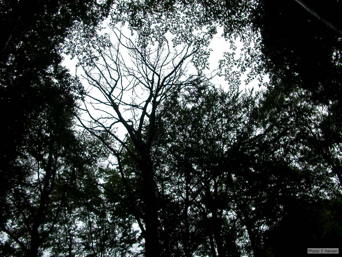 Dead beech in Germany |
P. cambivora colony morphology on PDA  Rosaceous colony morphology at 14 days at 20°C on PDA |
P. cambivora coralloid hyphae 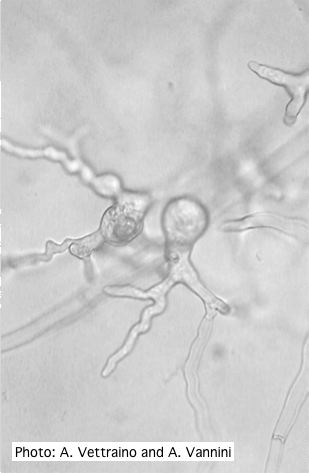 Coralloid hyphae with hyphal swelling-like structures |
|
P. cambivora colony morphology on PDA 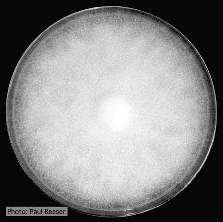 Colony morphology on PDA at 14 days |
P. cambivora colony morphology on PDA 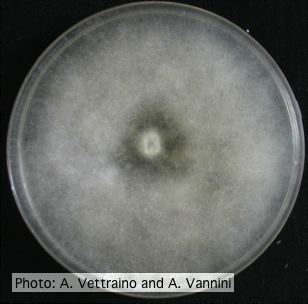 Uniform fluffy colony morphology at 14 days at 20°C on PDA |
P. cambivora sporangium with internal extended proliferation 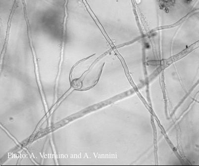 Empty sporangia showing internal extended proliferation |