

Sporangium with internal proliferation, photo from Q-bank, used with permission.
Photo Gallery
|
P. pinifolia sporangia 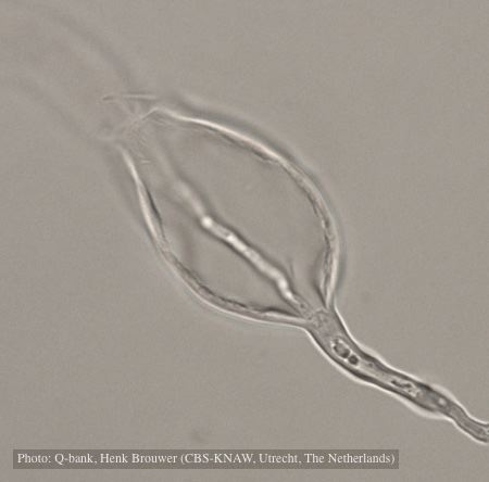 |
P. cryptogea sporangia  Sporangiophore showing internal proliferation through empty sporangia after zoospore release |
Chlamydospore of P. lateralis 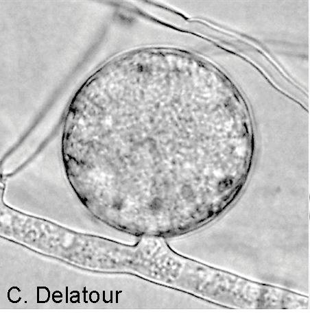 Terminal chlamydospore on a short side stalk |
|
P. austrocedrae hyphal swellings in liquid media drawing  Morphology of hyphae of Phytophthora austrocedrae, from Greslebin et al. 2007 |
P. alni sporangia 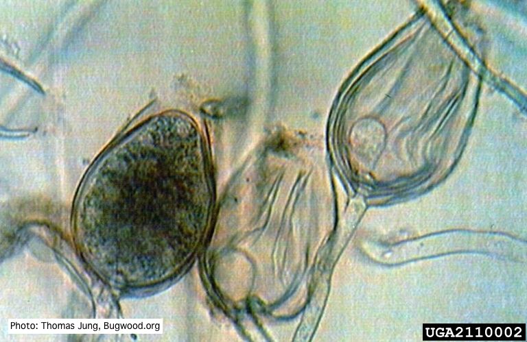 Non-papillate sporangia of P. alni showing nested proliferation. |
P. alni in riparian alder, Scotland 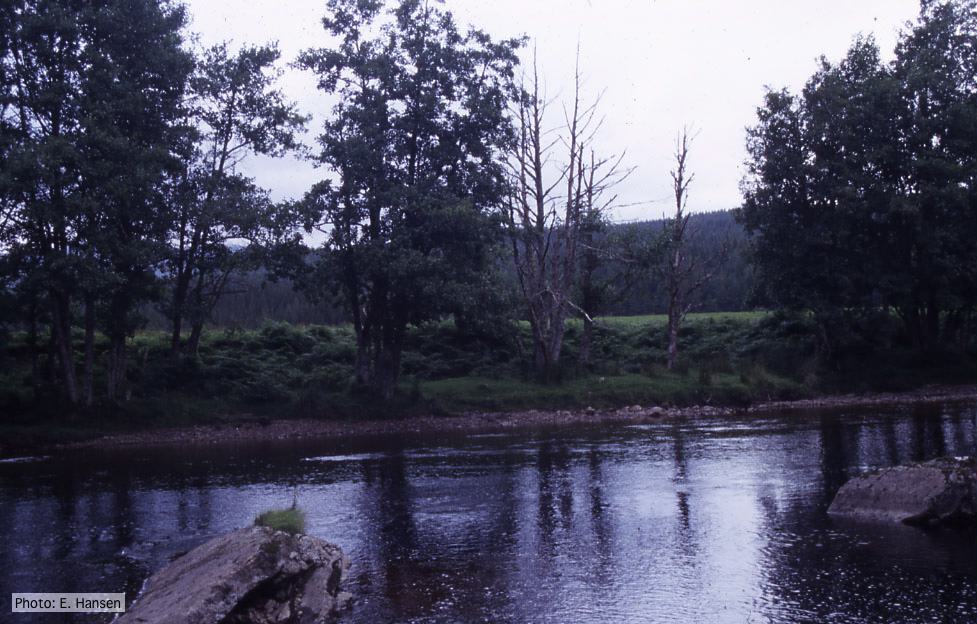 P. alni in riparian alder, Scotland |
|
P. cinnamomi hyphal swelling 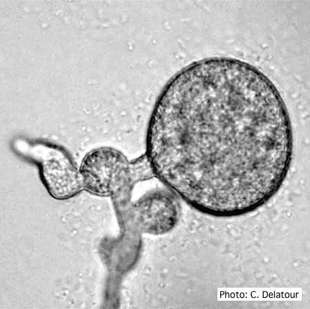 P. cinnamomi hyphal swelling (or thin walled chlamydospores) |
P. ramorum colony morphology on CMA PARP 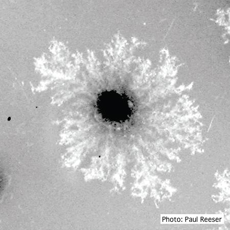 P. ramorum colony morphology on CMA PARP |
P. pinifolia coenocytic hyphae 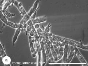 Coenocytic hyphae (from Duran et al. 2008). Scale bar = 20 μm. |
|
P. katsurae disease symptoms  Infected chestnut tree with girdling canker on stem |
P. pinifolia on Pinus radiata 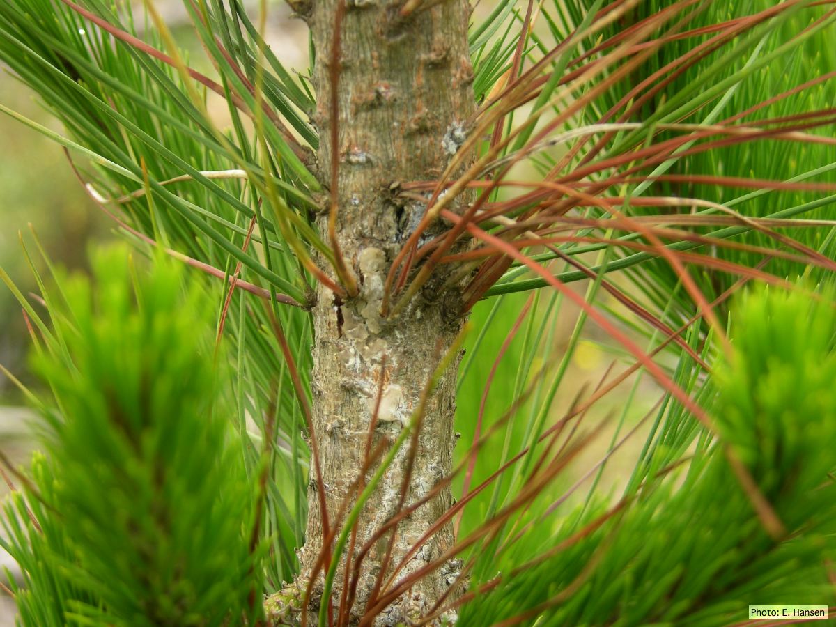 Pinus radiata, note Stem canker associated with necrotic needles. |
Growth of P. megakarya on V8 agar 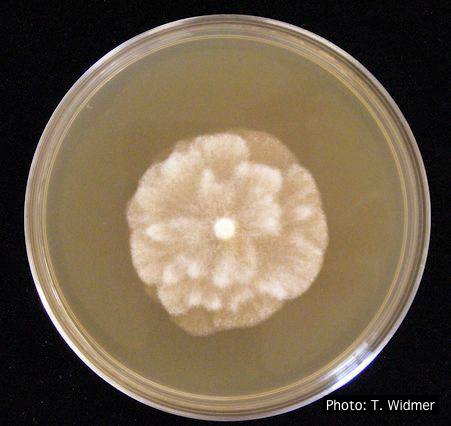 Growth of P. megakarya on V8 agar |