

P. cinnamomi colony growth on V8 at 14 days
Photo Gallery
|
P. cinnamomi colony morphology on V8 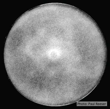 |
P. tentaculata hyphae 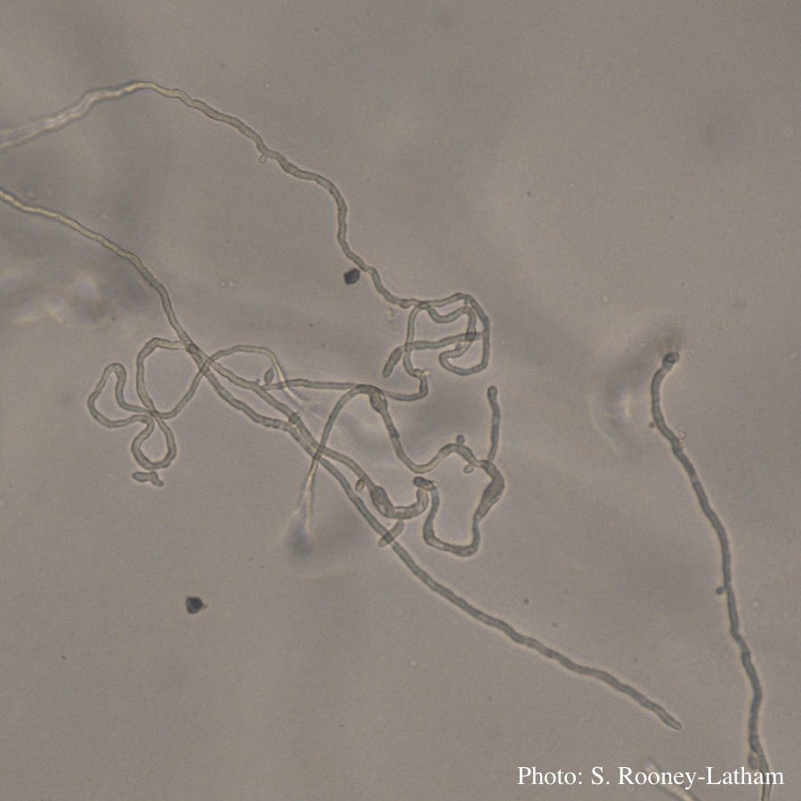 Looping hyphae commonly seen with P. tentaculata on PARP media |
P. alni subsp alni sporangium 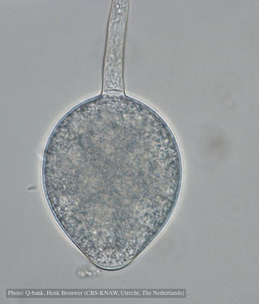 Non-papillate, non caducous sporangium, photo used with permission from Q-bank |
|
P. lateralis on Port Orford cedar  Typical decline of Chaemacyparis lawsoniana in Landrévarzec, France |
P. siskiyouensis bleeding canker  Bole lesions in the tissues under the bark of a bleeding canker: discoloration in the secondary phloem tissue |
P. austrocedrae hyphal swellings in liquid media drawing  Morphology of hyphae of Phytophthora austrocedrae, from Greslebin et al. 2007 |
|
Growth morphology on V8 of P. lateralis 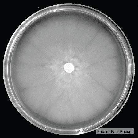 Colony morphology on V8 at 14 days |
P. austrocedrae - Mal del ciprés, stages of decline 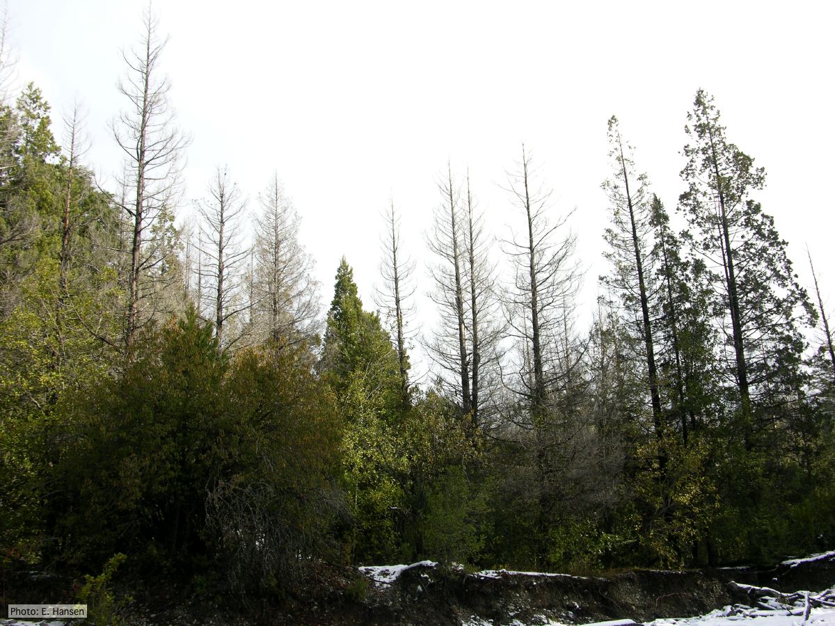 Colony morphology of P. austrocedrae at 16 C after four weeks on PDA |
P. ramorum sporangium  Deciduous sporangium, photo from Q-bank, used with permission |
|
P. lateralis on Port Orford cedar 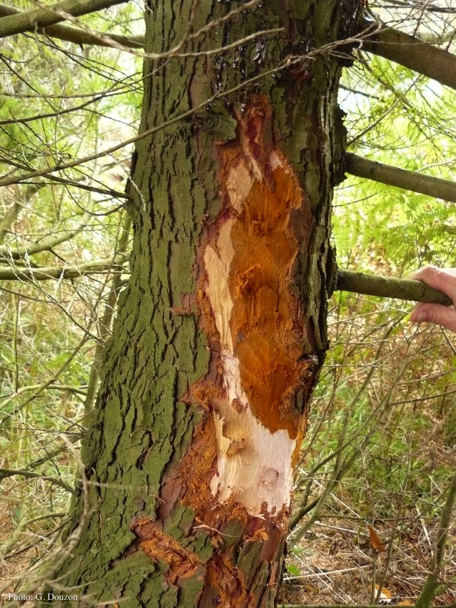 Bole lesion on Chaemacyparis lawsoniana in Lopérec, France |
P. chlamydospora chlamydospore  Phytophthora chlamydospora chlamydospore in agar. Bar is 20µm. |
P. kernoviae colony morphology on CMA PARPH 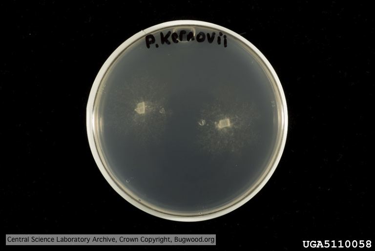 Organism grown on CMA PARP[H]; Plant disease 70, 1038-1043 |