

Rosacous colony morphology at 14 days at 20°C on MA
Photo Gallery
Site will be retired 9/1/2026
This site is no longer being developed and will be retired on September 1, 2026. Please contact us if you have any questions or would like to provide support to continue the project.
|
P. cambivora colony morphology on MA 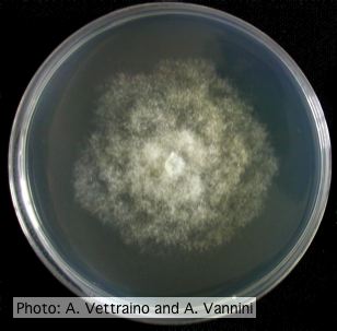 |
P. nicotianae colony morphology on PDA 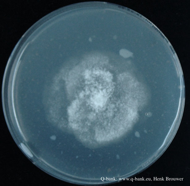 Phytophthora nicotianae CBS 321.49 PDA after 7 days at 24 degrees. Photo from Q-bank: www.q-bank.eu, Henk Brouwer (CBS-KNAW, Utrecht, The Netherlands) |
P. palmivora oogonia  P. palmivora oogonia with antheridia |
|
P. agathidicia oogonia 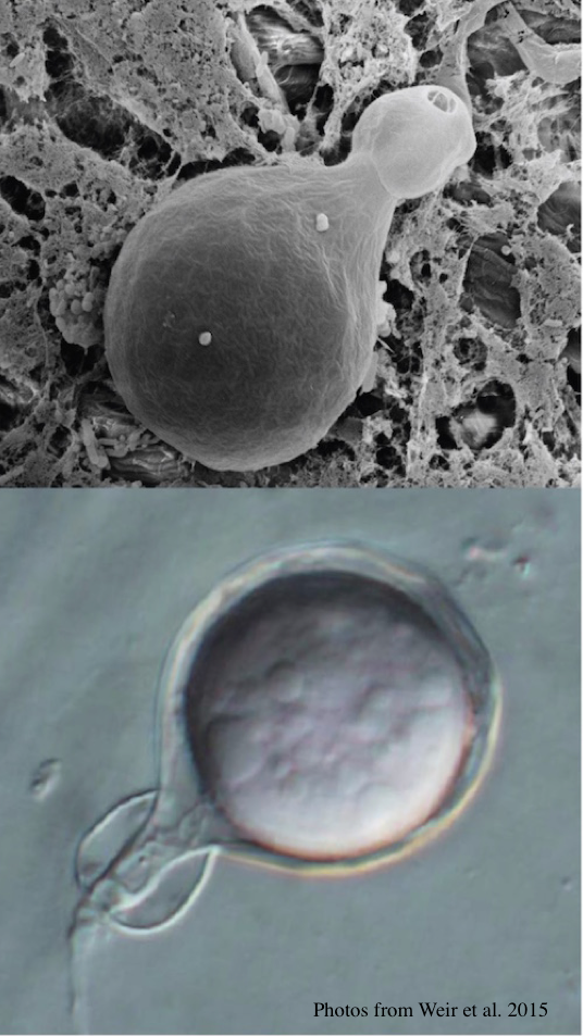 P. agathidicida oogonia with SEM (top) and light microscopy (bottom) |
Growth of P. megakarya on PDA 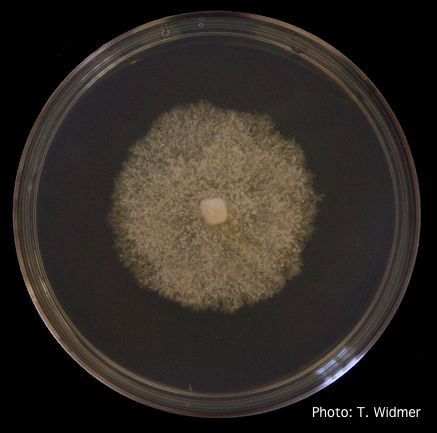 Growth of P. megakarya on potato dextrose agar |
Vehicle washing  Truck washing to avoid spread of P. lateralis |
|
P. pseudosyringae sporangium 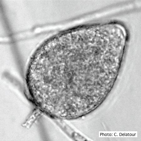 Ovoid, semipapillate sporangia showing medium length pedicel |
P. nicotianae colony morphology on V8 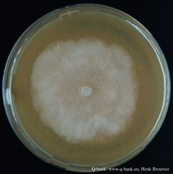 Phytophthora nicotianae CBS 321.49 V8 after 7 days at 24 degrees. Photo from Q-bank: www.q-bank.eu, Henk Brouwer (CBS-KNAW, Utrecht, The Netherlands) |
P. austrocedrae - Mal del ciprés in Argentina 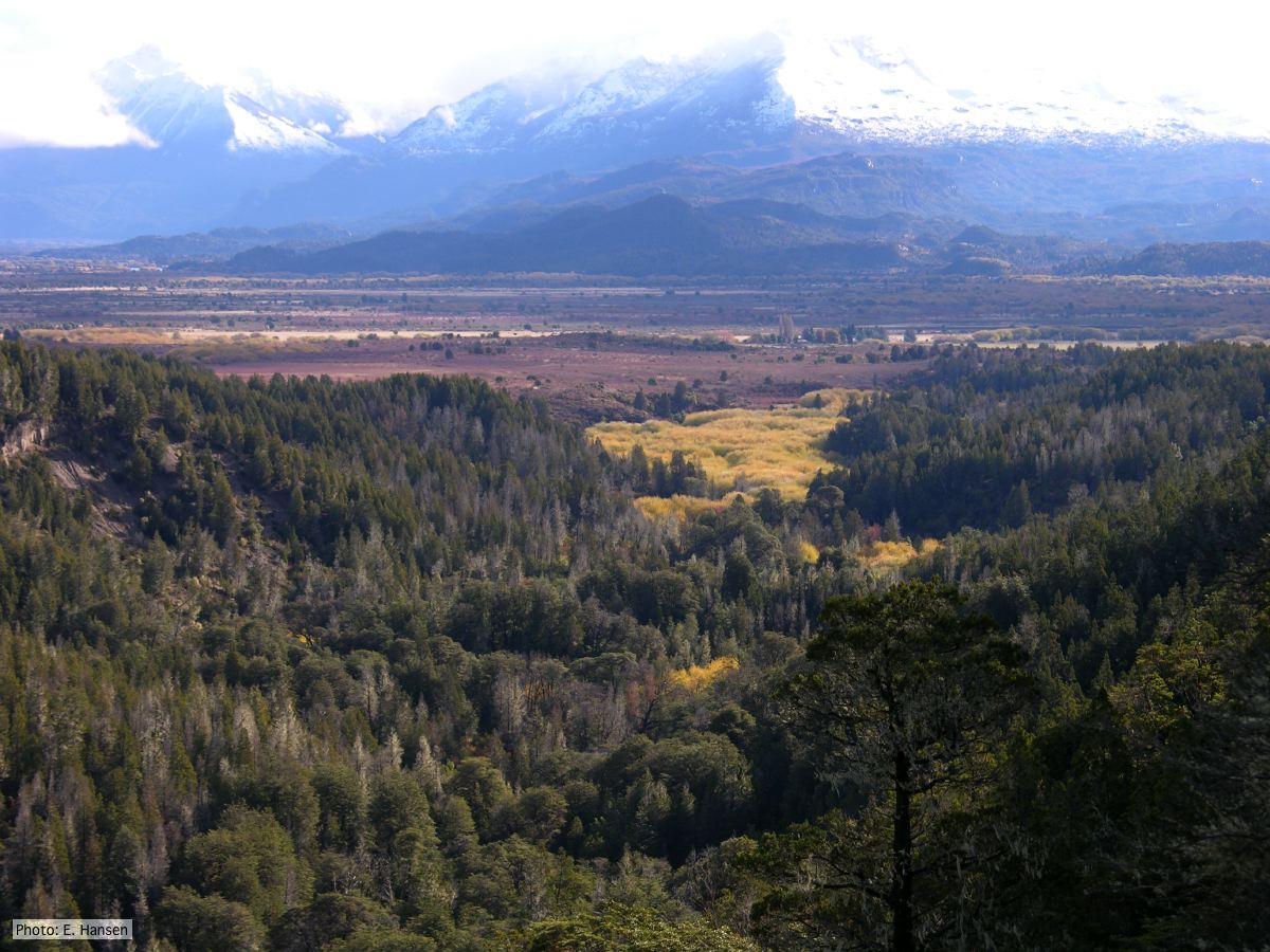 Mal del ciprés looking toward Rio Grande, Chubut Province, Argentina |
|
P. austrocedrae colony morphology on Tomato juice agar with B sitosterol 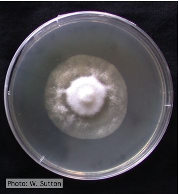 Colony morphology of P. austrocedrae at 16 ºC after 4 weeks on Tomato juice agar with B sitosterol |
P. arenaria hyphal swellings 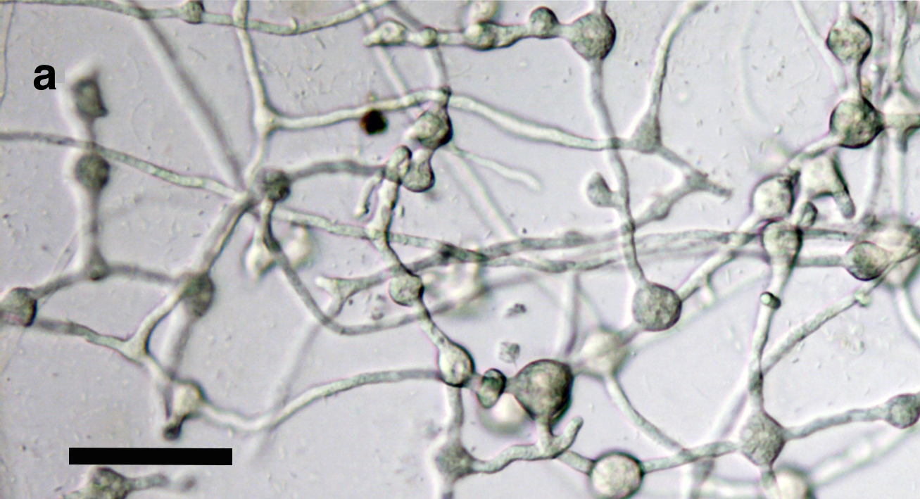 Catenulate, globose to subglobose hyphal swellings, some of them with radiating hyphae scale bar = 50 μm. |
P. pseudotsugae colony morphology on PDA  P. pseudotsugae colony growth on PDA agar |