

Phytophthora chlamydospora chlamydospore in agar. Bar is 20 µm.
Photo Gallery
Site will be retired 9/1/2026
This site is no longer being developed and will be retired on September 1, 2026. Please contact us if you have any questions or would like to provide support to continue the project.
|
P. chlamydospora chlamydospore 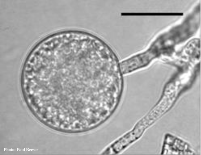 |
P. cambivora colony morphology on MA 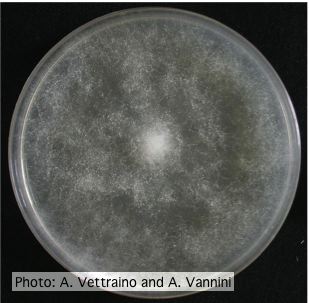 Uniform fluffy colony morphology at 14 days at 20°C on MA |
P. tentaculata oogonia and antheridia 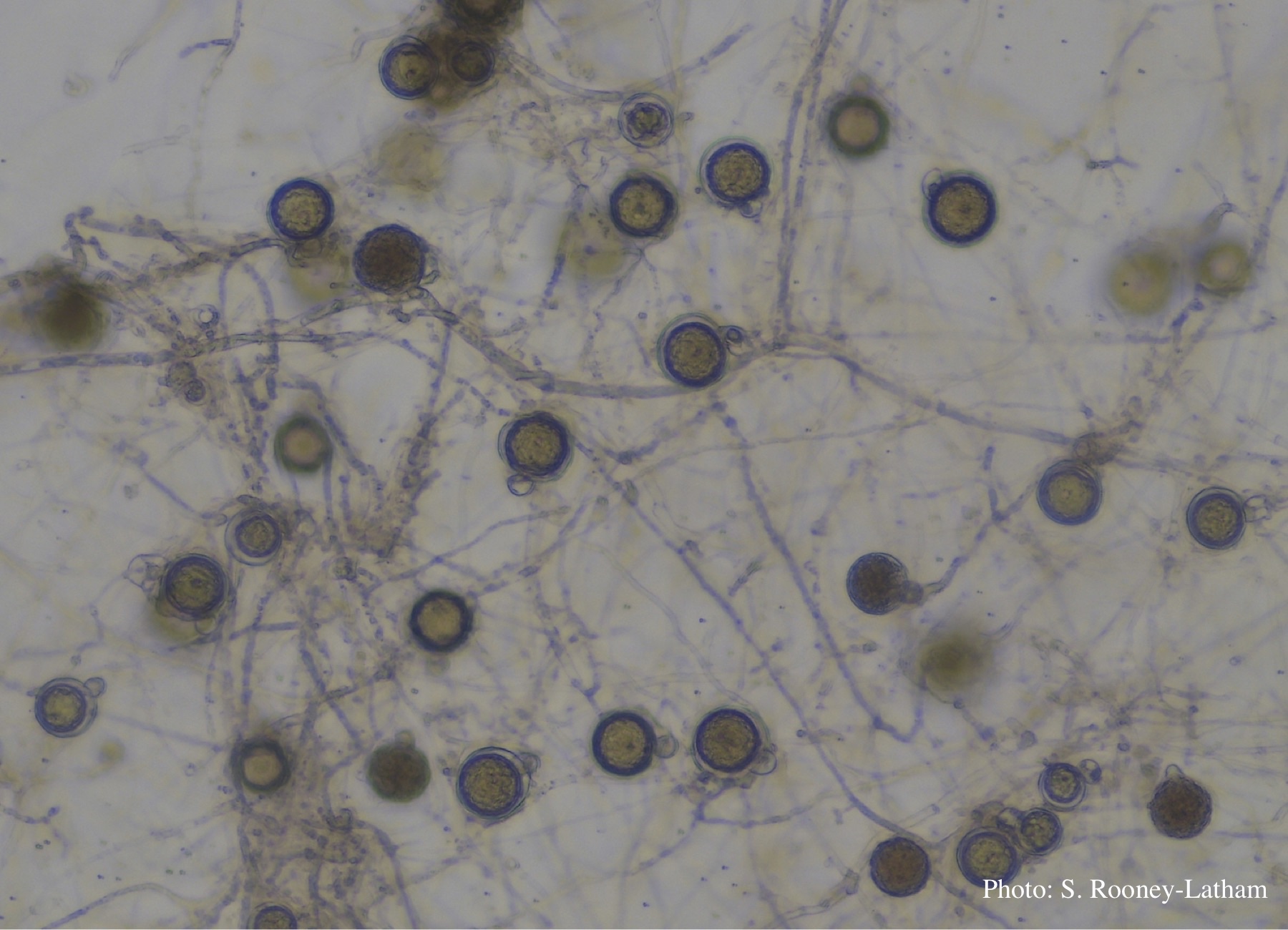 Oospores and oogonia with mostly paragynous but some amphigynous antheridia of P. tentaculata |
|
P. nicotianae sporangia  P. nicotianae overview of sporangia 40x. Photo from Q-bank: www.q-bank.eu, Henk Brouwer (CBS-KNAW, Utrecht, The Netherlands) |
P. chlamydospora chlamydospore 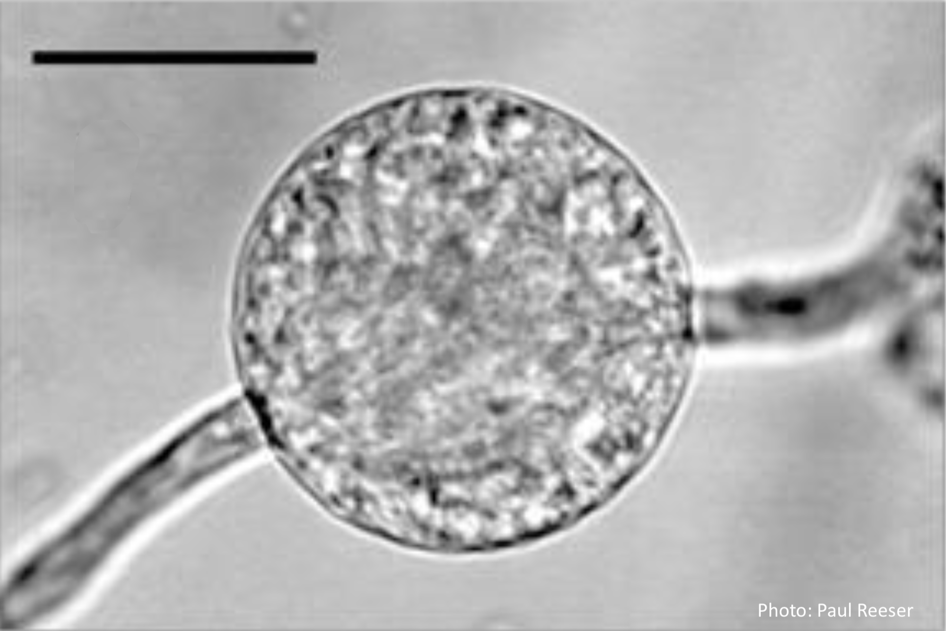 Phytophthora chlamydospora chlamydospore in agar. Bar is 20µm. |
P. tentaculata sporangium  Papillate sporangium of P. tentaculata |
|
Dying Port Orford Cedar trees 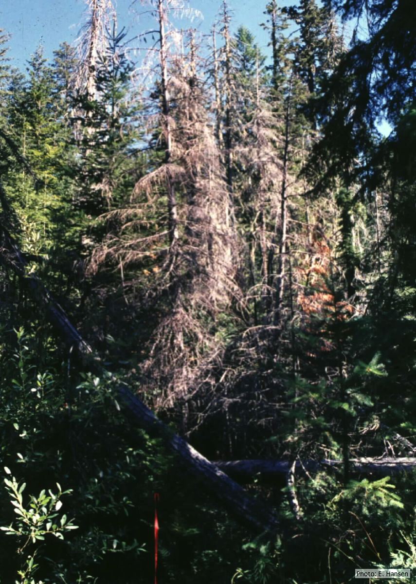 Dead Chamaecyparis lawsoniana trees |
Comparative gametangial morphology of Phytophthora Clade 5 species  Comparative gametangial morphology of Phytophthora Clade 5 species, with SEM (top) and light microscopy (bottom). P. heveae has smooth walled oogonia with funnel-shaped, amphigynous antheridia. P. agathidicida has mildly stipulate oogonia with globose amphigynous antheridia. P.cocois has mildly bullate oogonia with reflexed amphigynous antheridia. P. castaneae has coarsely bullate oogonium with rugose protuberances and narrow amphigynous antheridia (Weir et al. 2015). |
P. agathidicida lesion on kauri tree 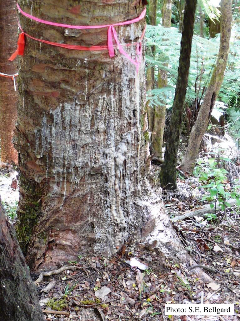 Advancing triangular lesion extending up the trunk of an 80 cm DBH kauri tree in the Huia Dam Site along Twin Peaks Track, Waitakere Regional Park |
|
P. palmivora sporangia, chlamydospores, hyphae 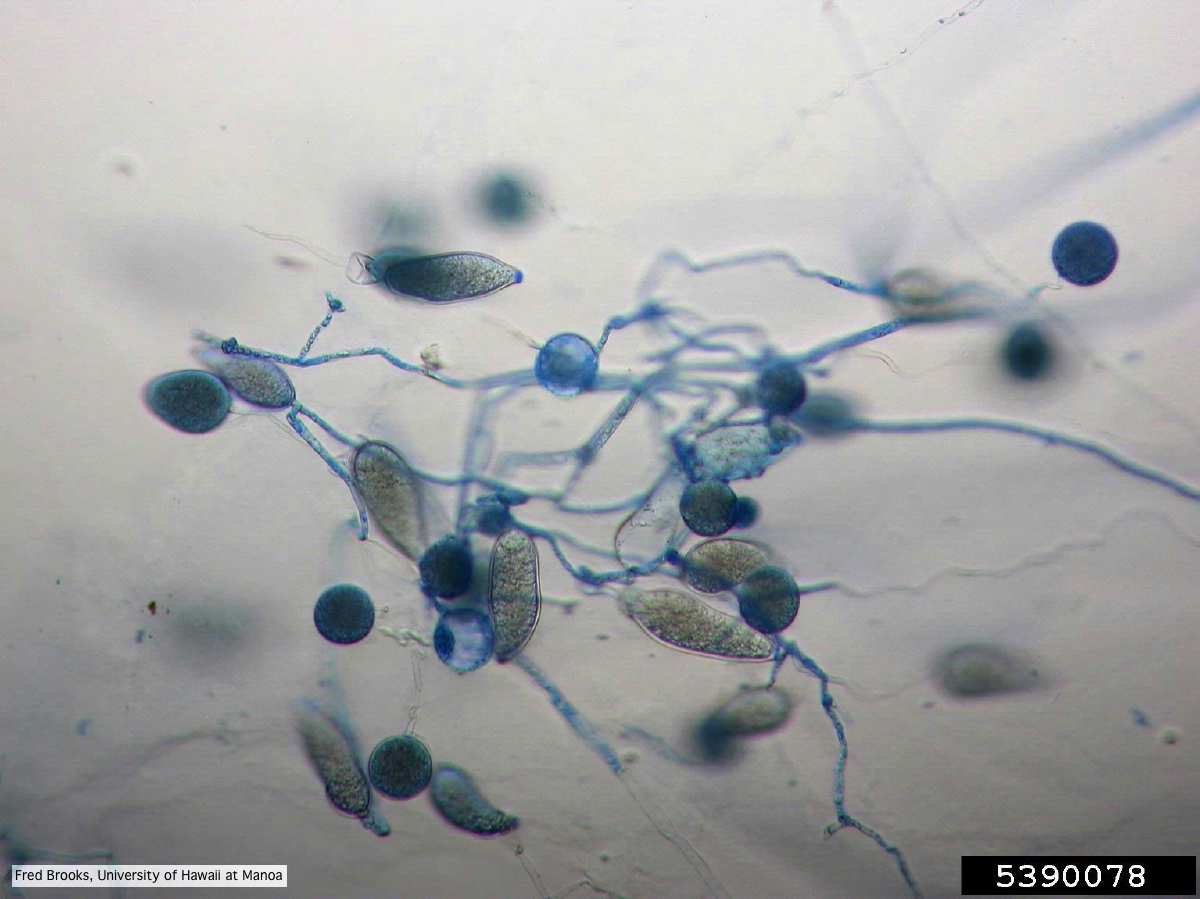 Sporangia (sporangiospores), chlamydospores, and hyphae stained with Cotton Blue |
P. pluvialis - appearance of new growth 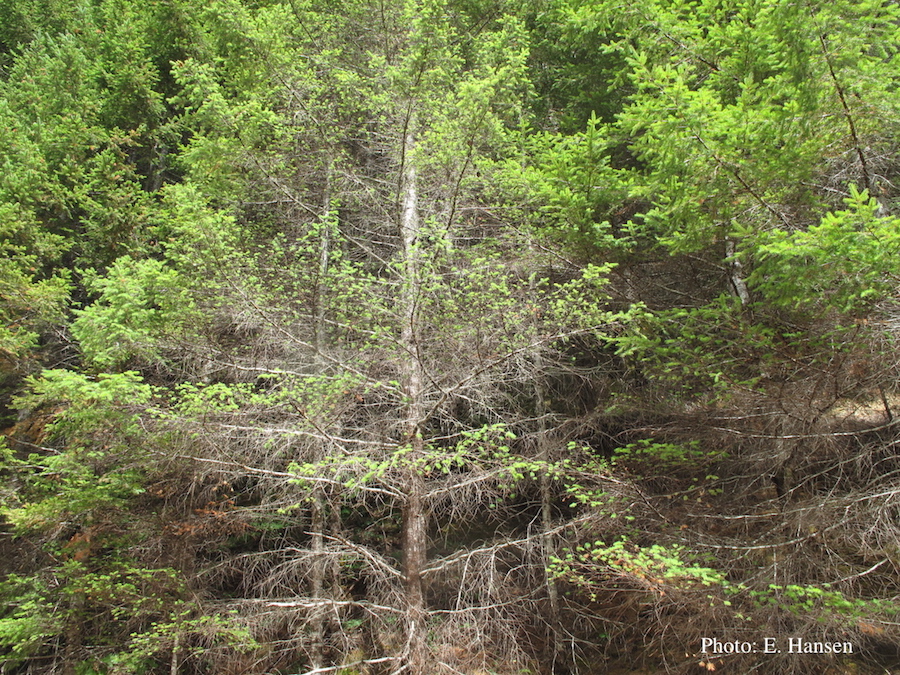 Tufted appearance of new growth from surviving buds on Douglas-fir, one year after defoliation. |
P. cambivora bole canker 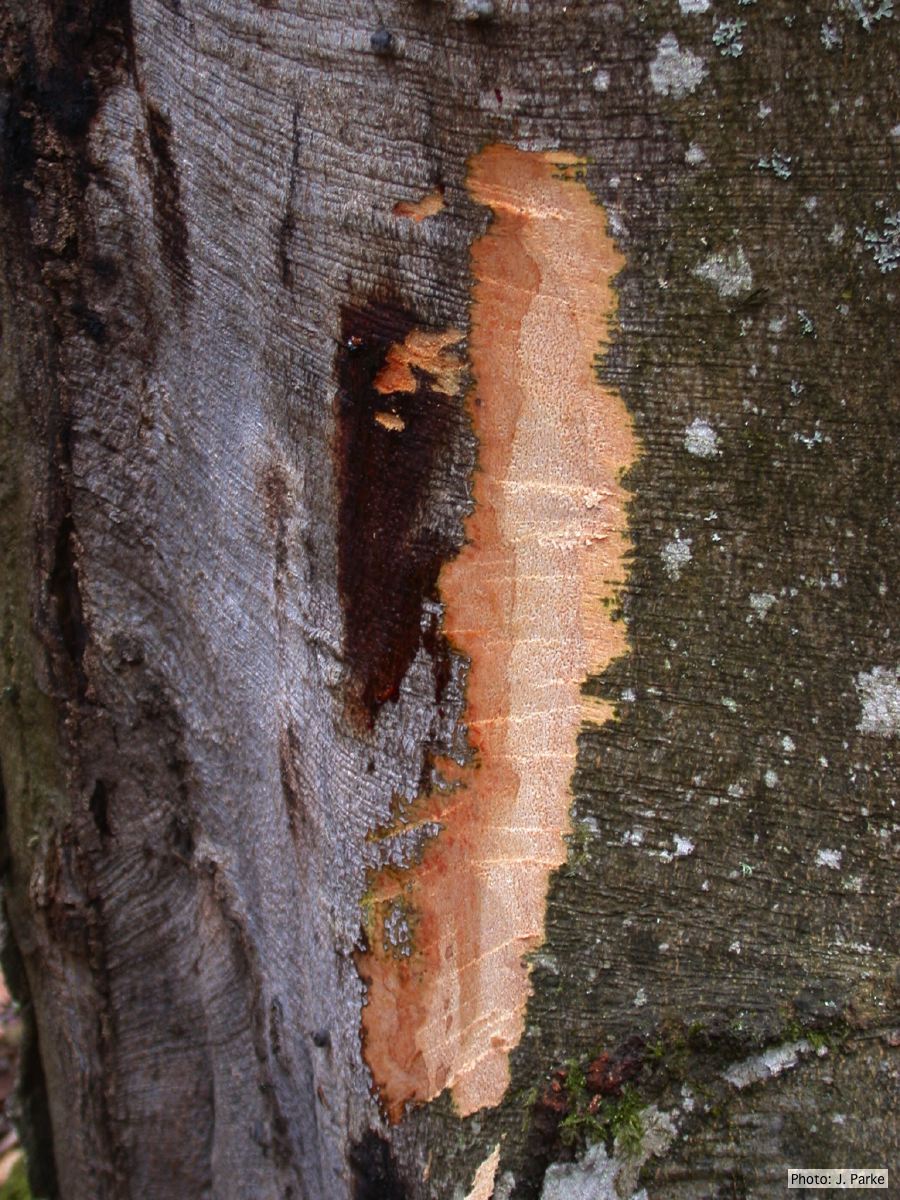 Fagus sylvatica bole canker |