

Colony morphology on V8 at 14 days
Photo Gallery
Site will be retired 9/1/2026
This site is no longer being developed and will be retired on September 1, 2026. Please contact us if you have any questions or would like to provide support to continue the project.
|
P. cryptogea colony morpholgy on V8  |
P. kernoviae sporangium 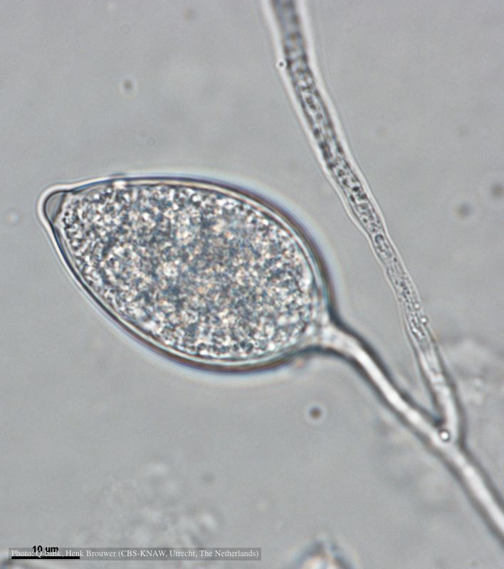 Papillate and caducous sporangium, photo from Q-bank, used with permission |
P. tentaculata microscopic characteristics 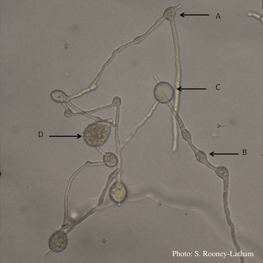 Hyphal swellings occuring at branching points of Mycelium (A), Intercalary hyphal swellings (B), Chlamydospore (C ), Sporangia (D) |
|
P. pinifolia colony morphology on V8 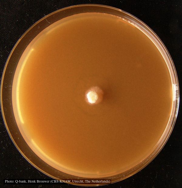 Colony pattern after 7 days on V8 at 24C, photo from Q-bank, used with permission |
Necrotic lesion in phloem caused by P. austrocedrae  Necrotic lesion in phloem with resin pocket caused by P. austrocedrae |
P. ramorum canker 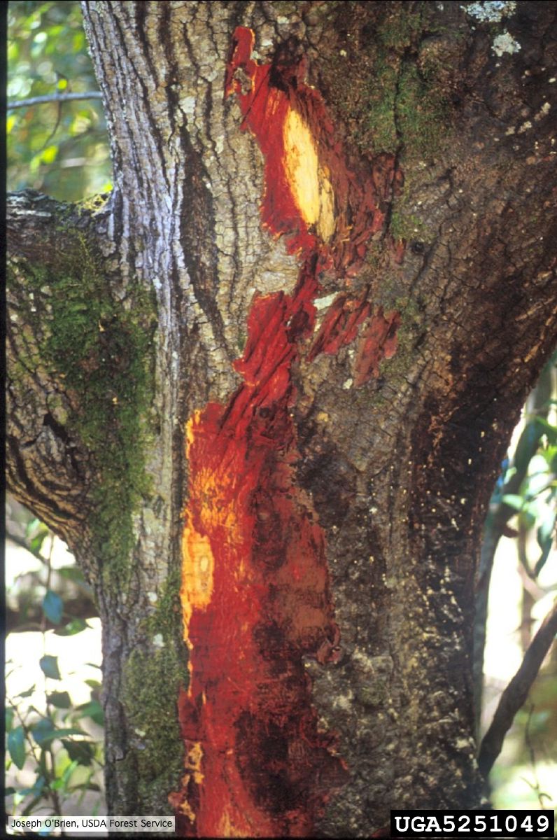 Bark discoloration and zone lines in coast live oak (Quercus agrifolia) |
|
P. frigida sporangia 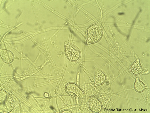 Noncaducous sporangia showing ovoid shape and papillate condition |
P. cinnamomi on Banksia 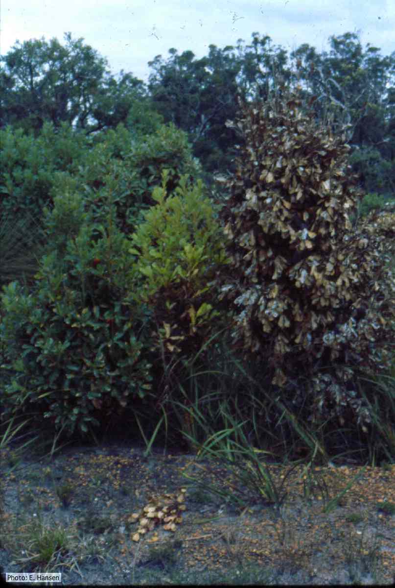 Dead and dying Banksia, Western Australia |
P. cambivora colony morphology on MA  Appressed colony morphology at 14 days at 20°C on MA |
|
P. cambivora oogonium 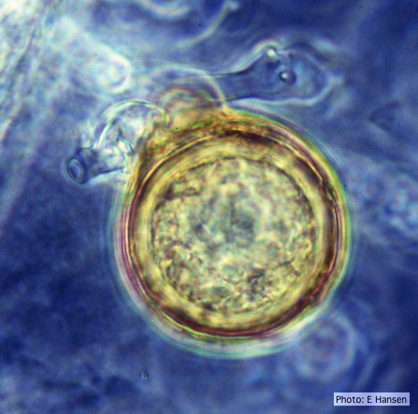 P. cambivora oogonium with antheridium |
P. pinifolia on Pinus radiata  Pinus radiata, note Stem canker associated with necrotic needles. |
P. tentaculata hyphae 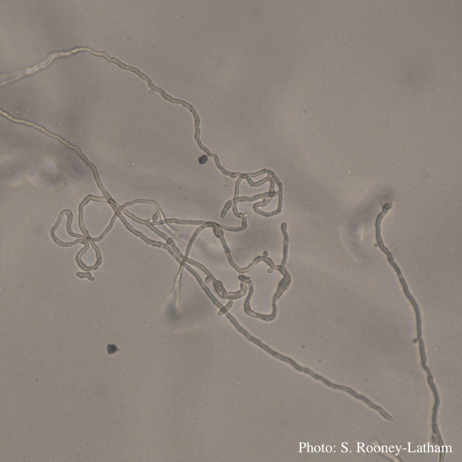 Looping hyphae commonly seen with P. tentaculata on PARP media |