

Diffuse, non-patterned, colony morphology of ICMP 16471 (the original “Gadgil isolate”) after 10-days incubation at 20°C in the dark
Photo Gallery
Site will be retired 9/1/2026
This site is no longer being developed and will be retired on September 1, 2026. Please contact us if you have any questions or would like to provide support to continue the project.
|
P. agathidicida growth on CMA 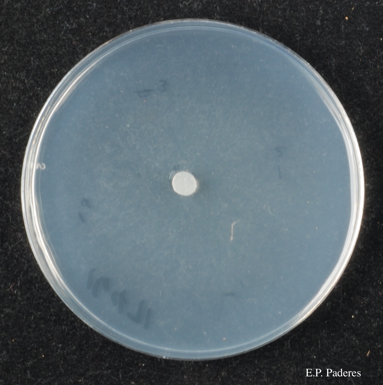 |
P. pluvialis symptoms on Douglas-fir 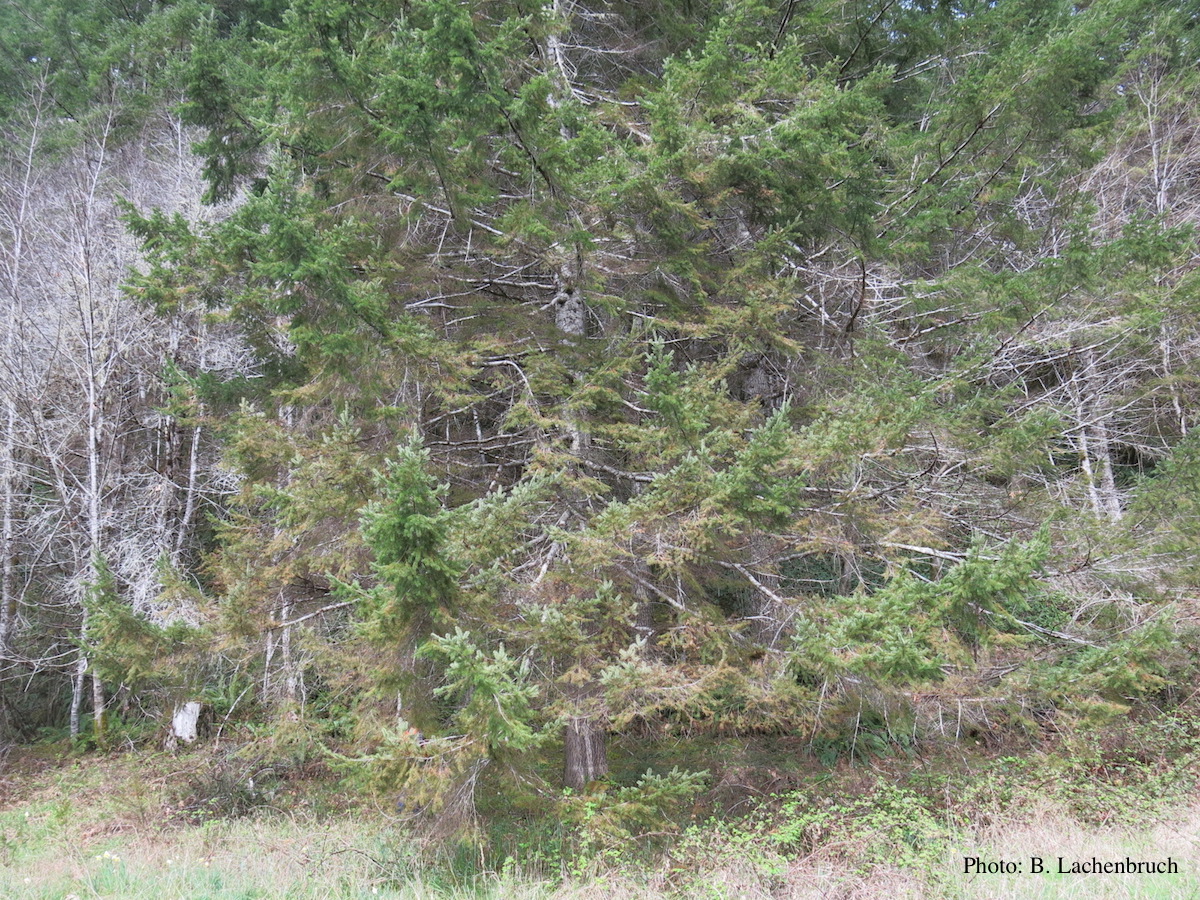 Red needle cast symptoms on Douglas-fir in western Oregon, 2015 |
P. cambivora colony morphology on PDA 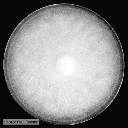 Colony morphology on PDA at 14 days |
|
P. agathidicia oogonia 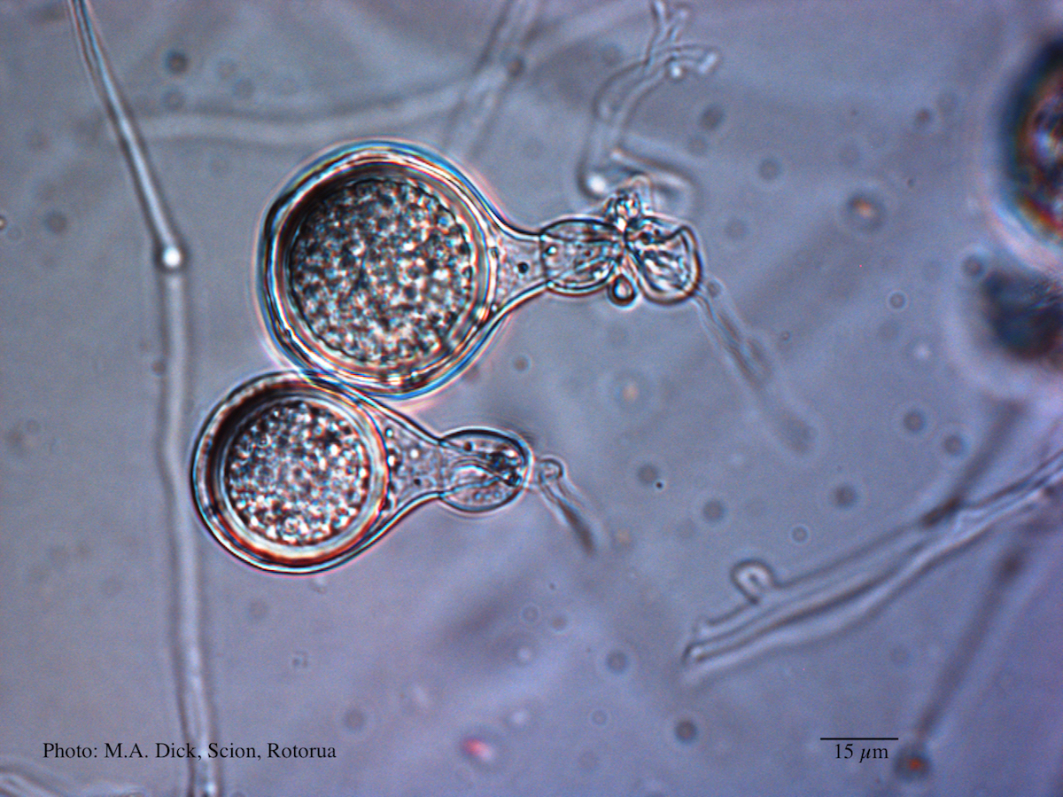 Light micrograph of P. agathidicida oospore (Scale bar equals 15 µm) |
P. pinifolia on Pinus radiata 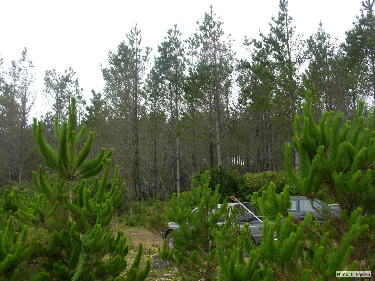 Pinus radiata stand, note Defoliation and regrowth |
P. pluvialis symptoms on Douglas-fir  Red needle cast symptoms on Douglas-fir in western Oregon, 2015 |
|
P. pinifolia on Pinus radiata 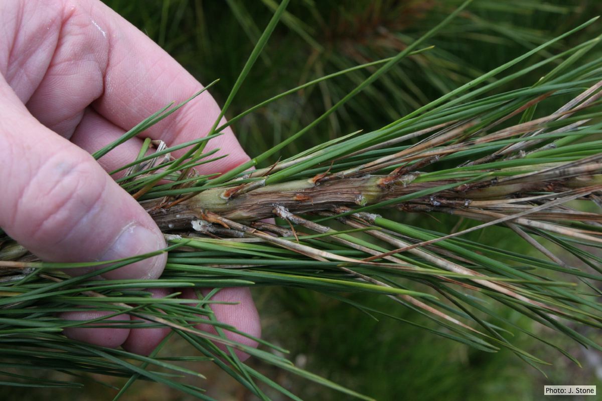 Pinus radiata needles, note “black line” symptom near needle bases |
P. kernoviae leaf wilt 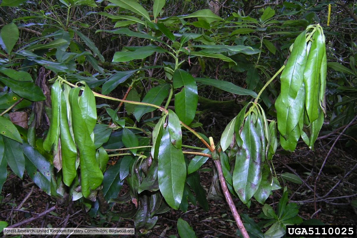 Wilted leaf of infected rhododendron |
P. cambivora colony morphology on PDA 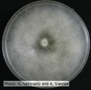 Uniform fluffy colony morphology at 14 days at 20°C on PDA |
|
P. pinifolia on Pinus radiata  Pinus radiata, note Stem canker associated with necrotic needles. |
P. siskiyouensis sporangia 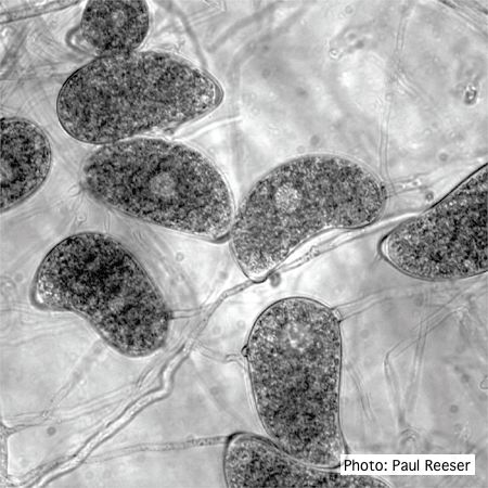 Sporangia showing a variety of shapes and orientations of semi-papillae and sporangiophores |
P. chlamydospora chlamydospore  Phytophthora chlamydospora chlamydospore in agar. Bar is 20µm. |