

Colony morphology at 7 days at 18°C on V8, photo from Q-bank, used with permission.
Photo Gallery
Site will be retired 9/1/2026
This site is no longer being developed and will be retired on September 1, 2026. Please contact us if you have any questions or would like to provide support to continue the project.
|
P. kernoviae colony morphology on V8 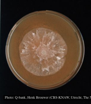 |
P. cambivora inactive lesion on chinquapin  Inactive lesion of P. cambivora on chinquapin |
P. chlamydospora chlamydospore 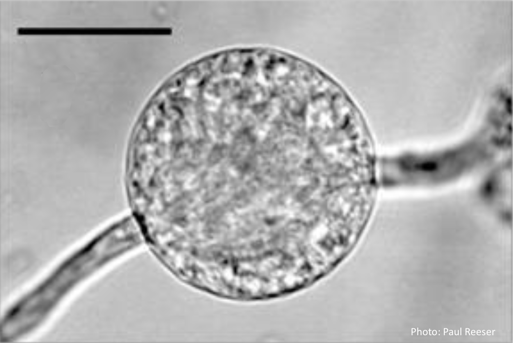 Phytophthora chlamydospora chlamydospore in agar. Bar is 20µm. |
|
Vehicle washing  Truck washing to avoid spread of P. lateralis |
P. alni oogonia 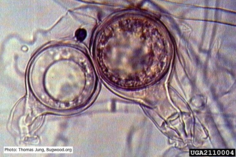 Smooth-walled oogonium of P. alni (Swedish variant) with oospore and amphigynous antheridium. |
P. pluvialis hyphal swellings 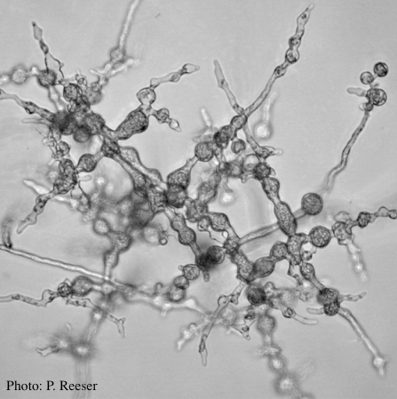 P. pluvialis hyphal swellings on agar |
|
P. pluvialis sporangia.  P. pluvialis sporangia on tape peel from infected Douglas-fir needle. |
P. palmivora colony morphology on PDA 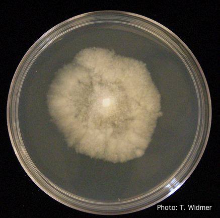 P. palmivora colony morphology on PDA |
P. cryptogea sporangium  Obpyriform non-papillate sporangia in water |
|
P. cactorum sporangia 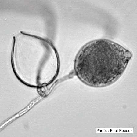 Broadly ovoid, papillate sporangia in water. |
P. cactorum bleeding canker 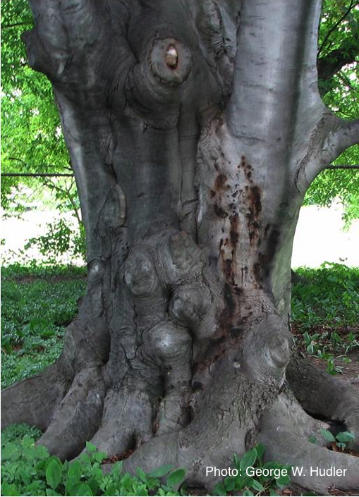 Bleeding canker on European beech (Fagus sylvatica) |
P. lateralis on Port Orford cedar  Atypical decline caused by aerial infections in Scaër, France |